ISSN 1004-4140
CN 11-3017/P
| Citation: |
JIN M, ZHAO X M, XU X, et al. Risk-Prediction Model Construction of Visceral Pleural Invasion in Early Lung Adenocarcinoma Based on Computed Tomography Imaging Features[J]. CT Theory and Applications, 2024, 33(4): 479-486. DOI: 10.15953/j.ctta.2023.107. (in Chinese).

|
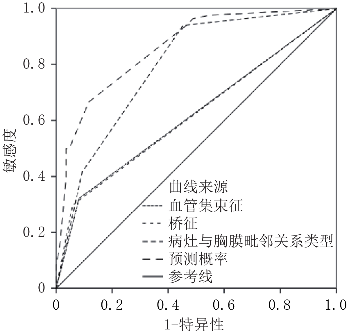
Objective: To construct a structural risk prediction model for visceral pleural invasion in early lung adenocarcinoma based on computed tomography (CT) features. Methods: 170 patients with early lung adenocarcinoma treated surgically in our hospital were retrospectively selected between January 2016 and December 2022 and grouped according into invasion (84 cases) and non-invasion (86 cases) groups. Independent risk factors related to CT imaging features of visceral pleural invasion in early lung adenocarcinoma were evaluated using univariate and multivariate factor methods. The effectiveness of the CT imaging feature prediction model for visceral pleural invasion in early subpleural lung adenocarcinoma was analyzed. Results: Univariate analysis showed that the proportion and maximum diameter of solid components, bridging sign, vascular bundle sign, and relationship between the lesion and pleura may be related to visceral pleural invasion in patients with early subpleural lung adenocarcinoma. Multivariate analysis showed that the bridging sign, vascular bundle sign, and type II/III relationship between the lesion and pleura were independent risk factors for visceral pleural invasion in early subpleural lung adenocarcinoma. The best cutoff values for predicting visceral pleural invasion in early subpleural lung adenocarcinoma using the bridge sign, vascular bundle sign, adjacent relationship between the lesion and pleura, and logistic model prediction probability were 0.50, 0.50, 0.50, and 56.25%, respectively. The Jordan index for each of these was 24.00%, 23.95%, 48.70%, and 55.04%, respectively. Conclusion: Based on the bridge sign, vascular cluster sign, and the relationship between tumor and pleural adjacency, the CT imaging feature model can be used to identify high-risk groups for visceral pleural invasion in early lung adenocarcinoma.
| [1] |
SUNG H, FERLAY J, SIEGEL R L, et al. Global cancer statistics 2020: Globocan estimates of incidence and mortality worldwide for 36 cancers in 185 countries[J]. CA: A Cancer Journal for Clinicians, 2021, 71(3): 209−249. DOI: 10.3322/caac.21660.
|
| [2] |
CHOI H, KIM H, HONG W, et al. Prediction of visceral pleural invasion in lung cancer on CT: Deep learning model achieves a radiologist-level performance with adaptive sensitivity and specificity to clinical needs[J]. European Journal of Radiology, 2021, 31(5): 2866−2876. DOI: 10.1007/s00330-020-07431-2.
|
| [3] |
ZHANG T, ZHANG J T, LI W F, et al. Visceral pleural invasion in T1 tumors (≤3 cm), particularly T1a, in the eighth tumor-node-metastasis classification system for non-small cell lung cancer: A population-based study[J]. Journal of Thoracic Disease, 2019, 11(7): 2754−2762. DOI: 10.21037/jtd.2019.06.32.
|
| [4] |
WO Y, ZHAO Y, QIU T, et al. Impact of visceral pleural invasion on the association of extent of lymphadenectomy and survival in stage I non-small cell lung cance[J]. Cancer Medicine, 2019, 8(2): 669−678. DOI: 10.1002/cam4.1990.
|
| [5] |
ZUO Z, LI Y, PENG K, et al. CT texture analysis-based nomogram for the preoperative prediction of visceral pleural invasion in cT1N0M0 lung adenocarcinoma: An external validation cohort study[J]. Clinical Radiology, 2022, 77(3): e215−e221. DOI: 10.1016/j.crad.2021.11.008.
|
| [6] |
YUAN M, LIU J Y, ZHANG T, et al. Prognostic impact of the findings on thinsection computed tomography in stage I lung adenocarcinoma with visceral pleural invasion[J]. Scientific Reports, 2018, 8(1): 1−9.
|
| [7] |
ONODA H, HIGASHI M, MURAKAMI T, et al. Correlation between pleural tags on CT and visceral pleural invasion of peripheral lung cancer that does not appear touching the pleural surface[J]. European Journal of Radiology, 2021, 31(12): 9022−9029. DOI: 10.1007/s00330-021-07869-y.
|
| [8] |
WEI S H, ZHANG J M, SHI B, et al. The value of CT radiomics features to predict visceral pleural invasion in ≤3 cm peripheral type early non-small cell lung cancer[J]. Journal of X-ray Science and Technology, 2022, 30(6): 1115−1126. DOI: 10.3233/XST-221220.
|
| [9] |
ZHANG Y, KWON W, LEE H Y, et al. Imaging assessment of visceral pleural surface invasion by lung cancer: Comparison of CT and contrast-enhanced radial T1-weighted gradient echo 3-Tesla MRI[J]. Korean Journal of Radiology, 2021, 22(5): 829−839. DOI: 10.3348/kjr.2020.0955.
|
| [10] |
SHI J, LI F, YANG F, et al. The combination of computed tomography features and circulating tumor cells increases the surgical prediction of visceral pleural invasion in clinical T1N0M0 lung adenocarcinoma[J]. Translational Lung Cancer Research, 2021, 10(11): 4266−4280. DOI: 10.21037/tlcr-21-896.
|
| [11] |
TU Z, LI C, TIAN T, et al. A risk classification system predicting the cancer-specific survival for postoperative stage IB non-small-cell lung cancer patients without lymphovascular and visceral pleural invasion[J]. Lung Cancer, 2021, 161(11): 114−121.
|
| [12] |
YANG X, SUN F, CHEN L, et al. Prognostic value of visceral pleural invasion in non-small cell lung cancer: A propensity score matching study based on the SEER registry[J]. Journal Surgery Oncology, 2017, 116(3): 398−406. DOI: 10.1002/jso.24677.
|
| [13] |
YU Y, HUANG R, WANG P, et al. Sublobectomy versus lobectomy for long-term survival outcomes of early-stage non-small cell lung cancer with a tumor size ≤2 cm accompanied by visceral pleural invasion: A SEER population-based study[J]. Journal of Thoracic Disease, 2020, 12(3): 592−604. DOI: 10.21037/jtd.2019.12.121.
|
| [14] |
NAM J G, PARK S, PARK C M, et al. Histopathologic basis for a chest CT deep learning survival prediction model in patients with lung adenocarcinoma[J]. Radiology, 2022, 305(2): 441−451. DOI: 10.1148/radiol.213262.
|
| [15] |
汤敏, 孙丹丹, 尹柯, 等. 胸膜下肺腺癌脏层胸膜侵犯CT及临床风险因素[J]. 放射学实践, 2020, 35(10): 1243−1248.
TANG M, SUN D D, YIN K, et al. CT and clinical risk factors of visceral pleural invasion in subpleural lung adenocarcinoma[J]. Radiology Practice, 2020, 35(10): 1243−1248. (in Chinese).
|
| [16] |
YANG S, YANG L, TENG L, et al. Visceral pleural invasion by pulmonary adenocarcinoma ≤3 cm: The pathological correlation with pleural signs on computed tomography[J]. Journal of Thoracic Disease, 2018, 10(7): 3992−3999. DOI: 10.21037/jtd.2018.06.125.
|
| [17] |
WANG F, LI P, LI F. Nomogram for predicting the relationship between the extent of visceral pleural invasion and survival in non-small-cell lung cancer[J]. Canadian Respiratory Journal, 2021, 20(7): 8816860.
|
| [18] |
WANG Y, LYU D, ZHOU T, et al. Multivariate analysis based on the maximum standard unit value of 18F-fluorodeoxyglucose positron emission tomography/computed tomography and computed tomography features for preoperative predicting of visceral pleural invasion in patients with subpleural clinical stage IA peripheral lung adenocarcinoma[J]. Diagnostic and Interventional Radiology, 2023, 29(2): 379−389. DOI: 10.4274/dir.2023.222006.
|
| [19] |
KIM H J, CHO J Y, LEE Y J, et al. Clinical significance of pleural attachment and indentation of subsolid nodule lung cancer[J]. Cancer Research and Treatment, 2019, 51(4): 1540−1548. DOI: 10.4143/crt.2019.057.
|
| [20] |
HEIDINGER B H, SCHWARZ-NEMEC U, ANDERSON K R, et al. Visceral pleural invasion in pulmonary adenocarcinoma: Differences in CT patterns between solid and subsolid cancers[J]. Radiology-Cardiothoracic Imaging, 2019, 1(3): e190071. DOI: 10.1148/ryct.2019190071.
|
Supported by: Beijing Renhe Information Technology Co. Ltd 


 DownLoad: