ISSN 1004-4140
CN 11-3017/P

| Citation: |
LI M M, FU Y G, XIAO Y, et al. CT Radiomics Nomogram Prediction for Tumor Deposits and Prognosis in Colorectal Cancer[J]. CT Theory and Applications, xxxx, x(x): 1-10. DOI: 10.15953/j.ctta.2024.055. (in Chinese).

|
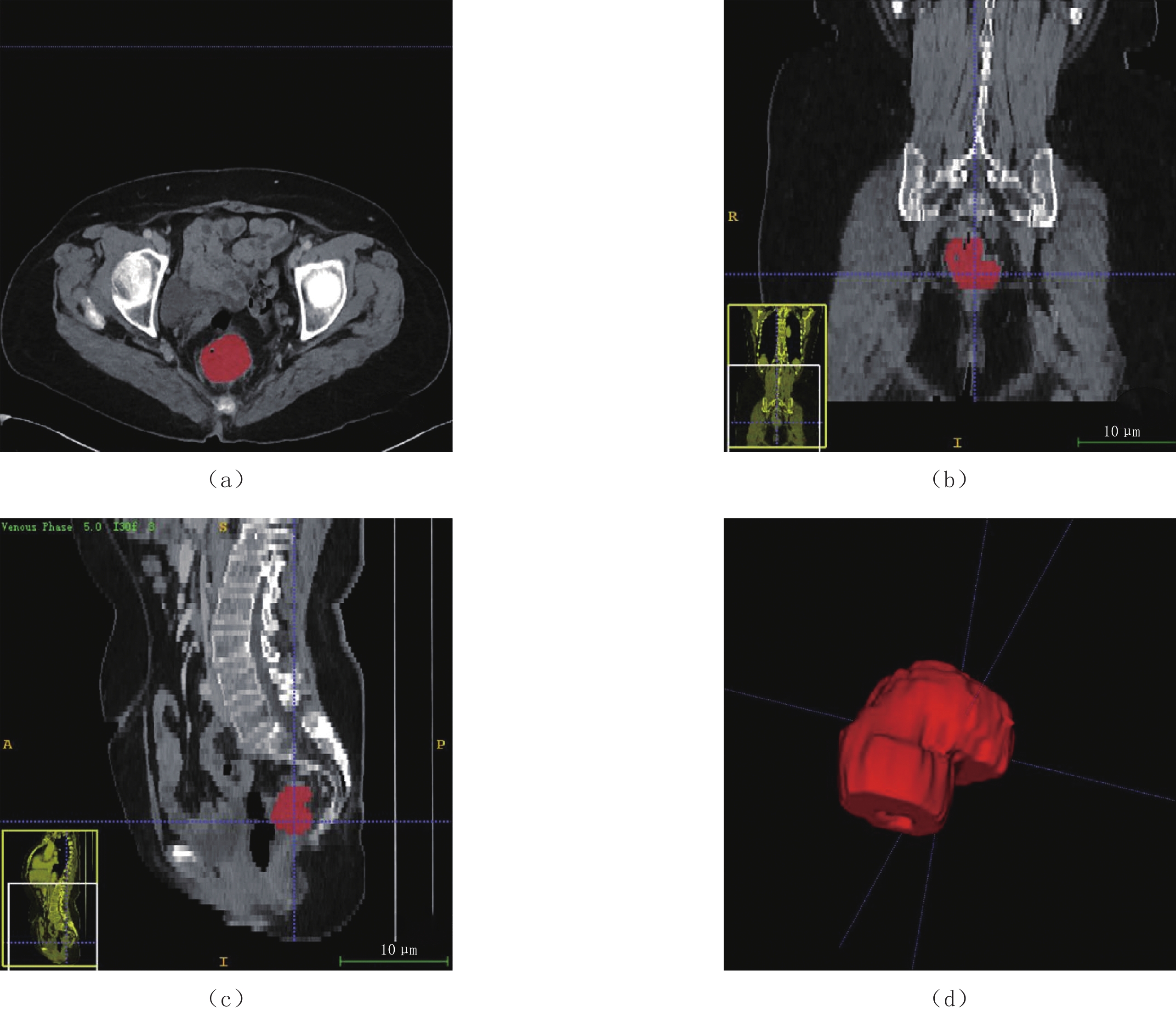
Objective: To establish CT radiomics nomogram for preoperative prediction of tumor deposits (TD) and recurrence-free survival (RFS) in patients with colorectal cancer (CRC). Methods: A retrospective study was conducted on 321 CRC patients confirmed by surgical pathology. The patients' data were divided were divided into a training set and a validation set at a ratio of 6:4, respectively. Radiomics features based on the primary tumor site were extracted from portal venous phase CT images, and the Least Absolute Shrinkage and Selection Operator (LASSO) algorithm was employed to select radiomics features associated with tumor deposits (TD). The least absolute shrinkage and selection operator (LASSO) regression algorithm was applied to choose radiomics features related to TD. A clinical-radiomics nomogram was developed based on the selected radiomics features and the most predictive clinical factors. Univariate and multivariate Cox regression analyses identified independent risk factors for a 3-year RFS. Results: The radiomics model achieved an area under the curve (AUC) of 0.80 in the training set and 0.79 in the validation set. By integrating radiomics features with clinical predictors (CEA, CA199, and CT-reported lymph node status), a nomogram was developed for the preoperative prediction of TD. The nomogram achieved an AUC of 0.85 in the training and validation sets. Furthermore, TD predicted by the nomogram was an independent risk factor for RFS, with poorer RFS observed in the TD-positive group compared to the TD-negative group. Conclusion: CT radiomics nomogram can effectively preoperatively predict TD and prognosis in CRC patients.
| [1] |
SIEGEL R L, MILLER K D, FUCHS H E, et al. Cancer statistics, 2022[J]. CA: A Cancer Journal for Clinicians, 2022, 72(1): 7−33. DOI: 10.3322/caac.21708.
|
| [2] |
RYU H S, KIM J, PARK Y R, et al. Recurrence patterns and risk factors after curative resection for colorectal cancer: Insights for postoperative surveillance strategies[J]. Cancers (Basel), 2023, 15(24): 5791. DOI: 10.3390/cancers15245791.
|
| [3] |
DELATTRE J F, Selcen Oguz Erdogan A, COHEN R, et al. A comprehensive overview of tumour deposits in colorectal cancer: Towards a next TNM classification[J]. Cancer Treatment Reviews, 2022, 103: 102325. DOI: 10.1016/j.ctrv.2021.102325.
|
| [4] |
MOON J Y, LEE M R, HA G W. Prognostic value of tumor deposits for long-term oncologic outcomes in patients with stage III colorectal cancer: a systematic review and meta-analysis[J]. International Journal of Colorectal Disease, 2022, 37(1): 141−151. DOI: 10.1007/s00384-021-04036-z.
|
| [5] |
PRICOLO V E, STEINGRIMSSON J, MCDUFFIE T J, et al. Tumor deposits in stage III colon cancer: Correlation with other histopathologic variables, prognostic value, and risk stratification-time to consider “N2c”[J]. American Journal of Clinical Oncology, 2020, 43(2): 133−138. DOI: 10.1097/COC.0000000000000645.
|
| [6] |
AMIN M B, GREENE F L, EDGE S B, et al. The eighth edition AJCC cancer staging manual: Continuing to build a bridge from a population-based to a more “personalized” approach to cancer staging[J]. CA: A Cancer Journal for Clinicians, 2017, 67(2): 93−99. DOI: 10.3322/caac.21388.
|
| [7] |
RATTO C, RICCI R, ROSSI C, et al. Mesorectal microfoci adversely affect the prognosis of patients with rectal cancer[J]. Diseases of the Colon and Rectum, 2002, 45(6): 733−743. DOI: 10.1007/s10350-004-6288-8.
|
| [8] |
ZHANG L N, XIAO W W, XI S Y, et al. Tumor deposits: Markers of poor prognosis in patients with locally advanced rectal cancer following neoadjuvant chemoradiotherapy[J]. Oncotarget, 2016, 7(5): 6335−6344. DOI: 10.18632/oncotarget.6656.
|
| [9] |
SHI M, ZHANG H, YAO G, et al. The role of tumor deposits in predicting the efficacy of chemotherapy in stage III colon cancer[J]. Frontiers in Oncology, 2020,10: 586603. Published 2020 Oct 14.
|
| [10] |
ROLLVEN E, ABRAHAM-NORDLING M, HOLM T, et al. Assessment and diagnostic accuracy of lymph node status to predict stage III colon cancer using computed tomography[J]. Cancer Imaging, 2017, 17(1): 3. DOI: 10.1186/s40644-016-0104-2.
|
| [11] |
ROLLVEN E, BLOMQVIST L, OISTAMO E, et al. Morphological predictors for lymph node metastases on computed tomography in colon cancer[J]. Abdominal Radiology (NY), 2019, 44(5): 1712−1721. DOI:10.1007/ s00261-019-01900-z.
|
| [12] |
CHEN L D, LI W, XIAN M F, et al. Preoperative prediction of tumour deposits in rectal cancer by an artificial neural network-based US radiomics model[J]. European Radiology, 2020, 30(4): 1969−1979. DOI: 10.1007/s00330-019-06558-1.
|
| [13] |
YANG Y S, FENG F, QIU Y J, et al. High-resolution MRI-based radiomics analysis to predict lymph node metastasis and tumor deposits respectively in rectal cancer[J]. Abdominal Radiology, 2021, 46(3): 873−884. DOI: 10.1007/s00261-020-02733-x.
|
| [14] |
MIZUNO H, MIYAKE H, NAGAI H, et al. Optimal cutoff value of preoperative CEA and CA19-9 for prognostic significance in patients with stage II/III colon cancer[J]. Langenbeck's Archives of Surgery, 2021, 406(6): 1987−1997. DOI: 10.1007/s00423-021-02236-3.
|
| [15] |
SHAN J, GU B, SHI L, et al. Prognostic value of CEA and CA19-9 in patients with local advanced rectal cancer receiving neoadjuvant chemoradiotherapy, radical surgery and postoperative chemotherapy[J]. Translational Cancer Research, 2021, 10(1): 88−98. DOI: 10.21037/tcr-20-2269.
|
| [16] |
LONG Q, XU Y, MA G, et al. Prognostic value of tumor deposit counts in patients with stage III colorectal cancer: A population-based study[J]. Journal of Investigative Surgery, 2022, 35(7): 1502−1509. DOI: 10.1080/08941939.2022.2069306.
|
| 1. |
丁朋,皮开荣,马云龙,杜兴忠. 孔间电磁波CT技术正演模型研究. 水利规划与设计. 2023(04): 71-76 .

| |
| 2. |
丁朋,芦安贵,邹宇杰. 孔间电磁波CT采集数据质量评价方法研究. 工程地球物理学报. 2023(02): 266-272 .
| |
| 3. |
张威,赵龙辉. 跨孔电磁波CT技术在水利工程地质岩溶探测中的应用. 陕西水利. 2023(06): 110-112 .
| |
| 4. |
江振寅,姜杰,郑军,田必林. 多孔对电磁波CT剖面拼接的影响因素及拼接方法. 工程地球物理学报. 2023(05): 667-675 .
| |
| 5. |
胡俊杰,徐洪苗,王鹏,段春龙. 基于三维可视化的跨孔电磁波CT在岩溶勘察方面的应用. 工程地球物理学报. 2022(04): 443-449 .
| |
| 6. |
杨国梁. 跨孔电磁波CT及剪切波测试方法在岩溶地质勘探中的应用. 江苏建筑. 2022(06): 126-130+143 .
|
Supported by: Beijing Renhe Information Technology Co. Ltd 


 DownLoad: