ISSN 1004-4140
CN 11-3017/P

| Citation: |
LI Y M, CHEN Weizhi.. Correlation between Carotid Artery Fat Density and Atherosclerosis[J]. CT Theory and Applications, xxxx, x(x): 1-8. DOI: 10.15953/j.ctta.2024.170. (in Chinese).

|
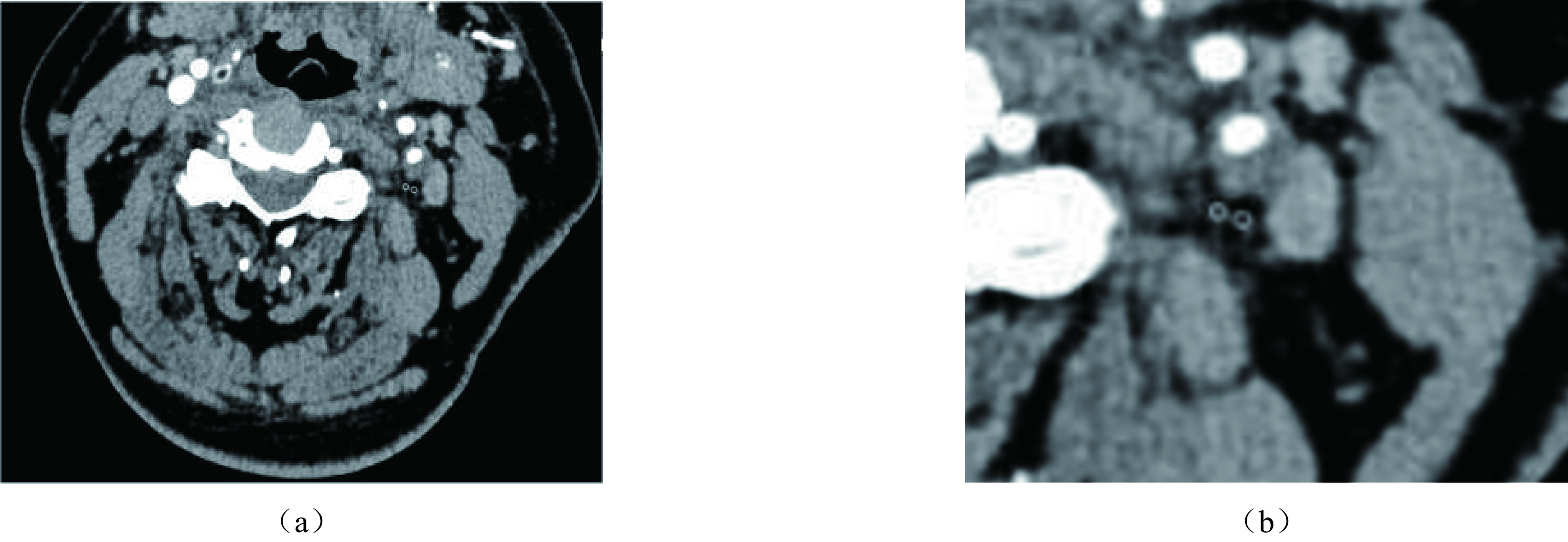
Objective: In this study, we aim to investigate the correlation between carotid artery fat density and atherosclerotic stenosis, providing a clinical reference for the early identification of carotid atherosclerotic stenosis. Methods: A retrospective analysis was conducted on patients with complete clinical data and confirmed diagnoses who underwent carotid computed tomography angiography (CTA) at Panjin Central Hospital between January 2023 and August 2023. A total of 224 blood vessels were included in the study. Patients were divided into a stenosis group and a control group based on the presence or absence of atherosclerosis. The stenosis group was further categorized into mild, moderate, and severe stenosis, and classified into Groups I, II, and III based on age. Additionally, patients were classified into symptomatic and asymptomatic groups based on their symptom status, and the correlation between perivascular fat density (PFD), carotid atherosclerotic stenosis, and cerebrovascular events was explored. Results: Hypertension, hyperlipidemia, and age were significantly associated with carotid atherosclerotic stenosis. PFD increased with the degree of stenosis and age. Correlation analysis revealed a positive correlation between PFD, age, history of hypertension, history of hyperlipidemia, and carotid artery stenosis. The PFD in the symptomatic group was higher than in the asymptomatic group, and the difference remained statistically significant after binary logistic correction for confounding factors. Conclusion: Carotid artery PFD is an independent risk factor for carotid atherosclerotic stenosis. Patients with symptomatic carotid artery stenosis exhibit higher carotid fat density compared to their asymptomatic counterparts.
| [1] |
LIU H H, JING J, WANG A X, ET AL. Stroke recurrence and antiplatelets in posterior versus anterior circulation minor stroke or transient ischemic attack[J]. Stroke, 2023, 54(4): 964-972. DOI: 10.1161/STROKEAHA.122.041738.
|
| [2] |
WAKSMAN R, MERDLER I, PATEL P, ET AL. Targeting inflammation in atherosclerosis: overview, strategy and directions[J]. EuroIntervention, 2024, 20(1): 32-44. DOI: 10.4244/EIJ-D-23-00606.
|
| [3] |
YUSUKE A, KAZUTAKA U, SEITARO N, ET AL. Beiging of perivascular adipose tissue regulates its inflammation and vascular remodeling[J]. Nature Communications, 2022, 13(1): 5117. DOI: 10.1038/s41467-022-32658-6.
|
| [4] |
QI X Y, QU S L, XIONG W H, ET AL. Perivascular adipose tissue (PVAT) in atherosclerosis: a double-edged sword[J]. Cardiovascular diabetology, 2018, 17(1): 1-20. DOI: 10.1186/s12933-017-0656-x.
|
| [5] |
CHENG C K, DING H, JIANG M, ET AL. Perivascular adipose tissue: Fine-tuner of vascular redox status and inflammation[J]. Redox Biology, 2023, 20(4): 102683.
|
| [6] |
冉值祯, 马跃, 侯阳. 心外膜脂肪组织密度对冠状动脉粥样硬化性心脏病的诊断价值[J]. 中国现代医学杂志, 2022, 32(1): 47-51. DOI: 10.3969/j.issn.1005-8982.2022.01.009.
RAN Z Z, MA Y, HOU Y. Diagnostic value of epicardial adipose tissue density in coronary atherosclerotic heart disease[J]. China Journal of Modern Medicine, 2022, 32(1): 47-51. DOI: 10.3969/j.issn.1005-8982.2022.01.009. (in Chinese).
|
| [7] |
MA R, VAN ASSEN M, SIDORENKOV G, ET AL. Relationships of pericoronary and epicardial fat measurements in male and female patients with and without coronary artery disease[J]. European Journal of Radiology, 2023, 169: 111154. DOI: 10.1016/j.ejrad.2023.111154.
|
| [8] |
MANUBOLU V S, LU J Y, MONTANO B, et al. Exploring the relationship between epicardial fat and coronary plaque burden and characteristics: Insights from cardiac CT imaging[J]. The International Journal of Cardiovascular Imaging. 2024, 40(9): 1951-1959.
|
| [9] |
孙万京, 仲玲珊, 李春阳, 等. 颈动脉分叉角同颈内动脉起始处粥样硬化的相关性[J]. 影像研究与医学应用, 2019, 3(7): 221. DOI: 10.3969/j.issn.2096-3807.2019.07.155.
SUN W J, ZHONG L S, LI C Y, ET AL. Correlation between carotid bifurcation Angle and atherosclerosis at the beginning of internal carotid artery[J]. Imaging Research and Medical Applicati on, 2019, 3(7): 221. DOI: 10.3969/j.issn.2096-3807.2019.07.155. (in Chinese).
|
| [10] |
余苗, 孟闫凯, 徐含波, 等. 颈动脉周围脂肪密度与急性缺血性脑卒中事件的相关性研究[J]. 临床放射学杂志, 2023, 42(6): 910-914.
YU M, MENG Y K, XU H B, ET AL. Correlation between peripheral carotid fat density and acute ischemic stroke[J]. Journal of Clinical Radiology, 2023, 42(6): 910-914. (in Chinese).
|
| [11] |
HEDIYEH B, K P M, PRANEIL P, ET AL. Association between carotid artery perivascular fat density and cerebrovascular ischemic events[J]. Journal of the American Heart Association, 2018, 7(24): 10383-10387.
|
| [12] |
中国心血管健康与疾病报告2022概要[J]. 中国循环杂志, 2023, 38(6): 583-612.
The Writing Committee of the Report on Cardiovascular Health and Diseases in China[J]. Chinese Circulation Journal, 2023, 38(6): 583-612. (in Chinese).
|
| [13] |
FLAHERTY M L, KISSELA B, KHOURY J C, ET AL. Carotid artery stenosis as a cause of stroke[J]. Neuroepidemiology, 2013, 40(1): 36-41. DOI: 10.1159/000341410.
|
| [14] |
CHENG S F, BROWN M M, SIMISTER R J, ET AL. Contemporary prevalence of carotid stenosis in patients presenting with ischaemic stroke[J]. The British Journal of Surgery, 2019, 106(7): 872-878. DOI: 10.1002/bjs.11136.
|
| [15] |
SONG P G, FANG Z, WANG H Y, ET AL. Global and regional prevalence, burden, and risk factors for carotid atherosclerosis: A systematic review, meta-analysis, and modelling study[J]. Lancet Glob Health, 2020, 8(5): 721-729. DOI: 10.1016/S2214-109X(20)30117-0.
|
| [16] |
AKIHIRO N, TOMOYO S, MAKOTO A, ET AL. Plaque Rupture, compared with plaque erosion, is associated with a higher level of pancoronary inflammation[J]. JACC. Cardiovascular imaging, 2021, 15(5): 828-839.
|
| [17] |
HARUHITO Y, TOMOYO S, KEISHI S, ET AL. Coronary inflammation and plaque vulnerability: A coronary computed tomography and optical coherence tomography study[J]. Circulation: Cardiovascular Imaging, 2023, 10(1): 59-63.
|
| [18] |
CASSIE H, I A G. The pathobiology of perivascular adipose tissue (PVAT), the fourth layer of the blood vessel wall[J]. Cardiovascular pathology: the official journal of the Society for Cardiovascular Pathology, 2022, 61(1): 459-461.
|
| [19] |
CAI M, ZHAO D, HAN X, ET AL. The role of perivascular adipose tissue-secreted adipocytokines in cardiovascular disease[J]. Frontiers in immunology, 2023, 14(1): 49-51.
|
| [20] |
RYSZARD N, J T G. Perivascular adipose tissue inflammation in vascular disease[J]. British journal of pharmacology, 2017, 174(20): 3496-3513. DOI: 10.1111/bph.13705.
|
| [21] |
BERMAN DS, KWIECINSKI J. Imaging coronary inflammatory risk[J]. JACC. Cardiovascular imaging, 2021, 15(3): 472-475.
|
| [22] |
OIKONOMOU E K, ANTONOPOULOS A S, SCHOTTLANDER D, ET AL. Standardized measurement of coronary inflammation using cardiovascular computed tomography: integration in clinical careas a prognostic medical device[J]. Cardiovascular research, 2021, 117(13): 2677-2690.
|
| [23] |
HU X, CHEN J, FU H, ET AL. Association between carotid artery perivascular fat density and embolic stroke of undetermined source[J]. Frontiers in Neurology, 2022, 12(1): 765962.
|
| [24] |
牛稳, 邱晓晖, 刘艺超. 颈动脉粥样硬化狭窄的血管周围脂肪密度与脑梗死之间的关系[J]. 实用医学杂志, 2023, 39(1): 103-108.
NIU W, QIU X H, LIU Y C. The relationship between perivascular fat density and cerebral infarction in carotid atherosclerosis[J]. Journal of Practical Medicine, 2023, 39(1): 103-108. (in Chinese).
|
| [25] |
SABA L, ZUCCA S, GUPTA A, ET AL. Perivascular fat density and contrast plaque enhancement: does a correlation exist[J]. AJNR Am J Neuroradiol, 2020, 41(8): 1460-1465 DOI: 10.3174/ajnr.A6710.
|
| [26] |
ZHANG D H, JIN J L, ZHU C F, ET AL. Association between carotid artery perivascular fat density and cerebral small vessel disease[J]. Aging, 2021, 13(14): 839-851.
|
| [27] |
HENRICHOT E, JUGE-AUBRY C E, PERNIN A, ET AL. Production of chemokines by perivascular adipose tissue: A role in the pathogenesis of atherosclerosis[J]. Arteriosclerosis, Thrombosis, and Vascular Biology, 2005, 25(12): 2594-2599. DOI: 10.1161/01.ATV.0000188508.40052.35.
|
| [28] |
USUI E, MATSUMURA M, MINTZ G S, ET AL. Clinical outcomes of low-intensity area without attenuation and cholesterol crystals in non-culprit lesions assessed by optical coherence tomography[J]. Atherosclerosis, 2021, 332(1): 41-47.
|
| [29] |
OIKONOMOU E K, MARWAN M, DESAI M Y, ET AL. Non-invasive detection of coronary inflammation using computed tomography and prediction of residual cardiovascular risk (the CRISP CT study): a post-hoc analysis of prospective outcome data[J]. Lancet, 2018, 392(10151): 929-939. DOI: 10.1016/S0140-6736(18)31114-0.
|
| [30] |
应伟峰, 陈穹, 张莹, 等. 冠状动脉易损与非易损斑块的冠周与主动脉根部心外膜脂肪CT衰减指数比值差异[J]. 中国医学影像学杂志, 2023, 31(8): 818-823. DOI: 10.3969/j.issn.1005-5185.2023.08.005.
YING W F, CHEN Q, ZHANG Y, ET AL. The difference of CT attenuation index ratio between pericardial and epicardial fat in vulnerable and non-vulnerable coronary plaques[J]. Chinese Journal of Medical Imaging, 2023, 31(8): 818-823. DOI: 10.3969/j.issn.1005-5185.2023.08.005.
|
| [31] |
MUSHENKOVA N V, SUMMERHILL V I, ZHANG D, ET AL. Current advances in the diagnostic imaging of atherosclerosis: insights into the pathophysiology of vulnerable plaque[J]. International Journal of Molecular Sciences, 2020, 21(8): 2992. DOI: 10.3390/ijms21082992.
|
| [32] |
李静, 乔建民, 王俊奇, 等. 心外膜及心周脂肪体积与颈动脉粥样斑块的关系[J]. CT理论与应用研究, 2017, 26(6): 761-768.
LI J, QIAO J M, WANG J Q, ET AL. Relationship between epicardial and pericardiac fat volume and carotid atherosclerotic plaque[J]. CT Theoretical and Applied Research, 2017, 26(6): 761-768.
|
| [33] |
ANTONOPOULOS AS, SANNA F, SABHARWAL N, ET AL. Detecting human coronary inflammation by imaging perivascular fat[J]. Science Translational Medicine, 2017, 9(398): 2658. DOI: 10.1126/scitranslmed.aal2658.
|
| [34] |
BARADARAN H, MYNENI P K, PATEL P, ET AL. Association between carotid artery perivascular fat density and cerebrovascular ischemic events[J]. Journal of the American Heart Association, 2018, 7(24): e010383. DOI: 10.1161/JAHA.118.010383.
|
| [35] |
XIA R, FAN S, JIAN H, ET AL. Impact of carotid hemodynamics on carotid plaque location: Age difference[J]. Neuro endocrinology letters, 2023, 44(6): 399-409.
|
| 1. |
翟浩宇,陈雪,杨冰炜,王成磊,吴颖,崔慕瑶,吴佳禾,李卫东. 基于R语言的国家专利中药复方调治肺结节的用药规律研究. 中国医药导报. 2025(08): 1-7 .

| |
| 2. |
崔瑞芳,李仁廷,韩梦影. 李仁廷从“五脏失和,癌毒内聚”辨治肺癌. 中医学报. 2024(08): 1728-1732 .
| |
| 3. |
邓永军. 基于五行学说肝肺脾生克制化理论治疗肺结节. 中医研究. 2024(08): 24-27 .
|
Supported by: Beijing Renhe Information Technology Co. Ltd 


 DownLoad: