ISSN 1004-4140
CN 11-3017/P

| Citation: |
MENG X H, CUI X F, CHEN Y M, et al. Exploration of the Application Value of Dual Energy CT in the Diagnosis and Phase IIA Display of Talar Osteochondrial Injury[J]. CT Theory and Applications, xxxx, x(x): 1-9. DOI: 10.15953/j.ctta.2024.220. (in Chinese).

|
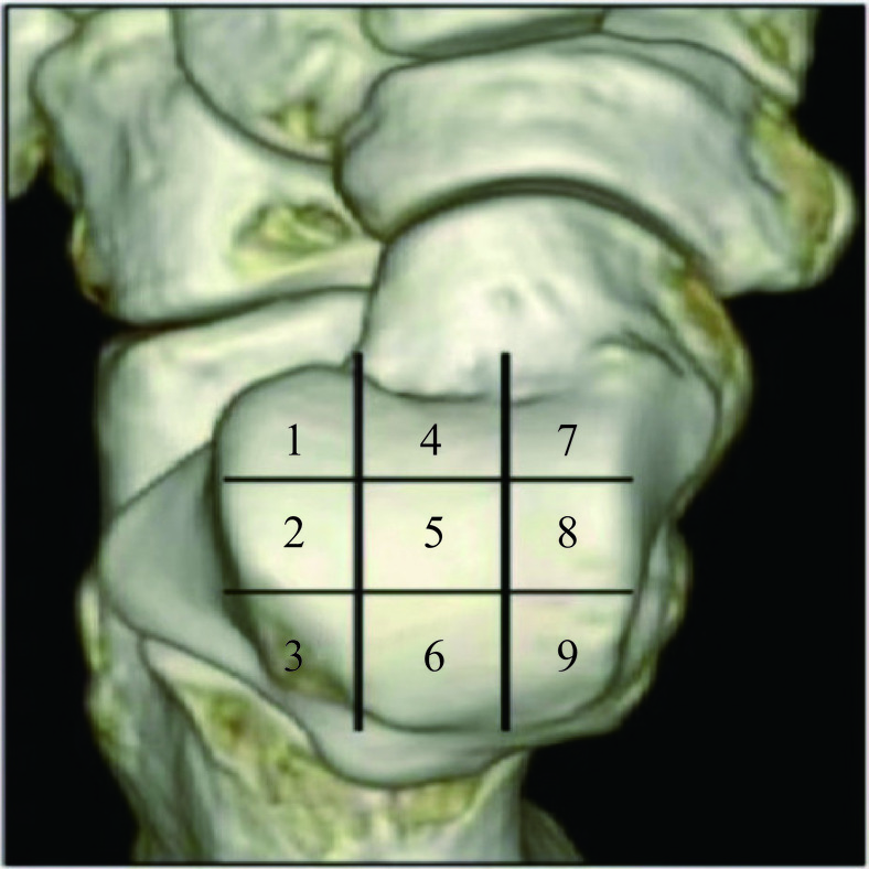
Objective: To investigate the diagnostic value of dual-energy computed tomography (DECT) for Stage IIA talar osteochondral injuries, with magnetic resonance imaging (MRI) as the reference standard. Methods: Thirty consecutive patients admitted to the orthopedics department of our hospital from January 2023 to December 2023, with long-term ankle pain and diagnosed with an osteochondral lesion of the talus through clinical and MRI examinations, were enrolled. DECT was performed on the patients within 24 hours after the MRI examination. Doctor A diagnosed and staged the 30 MRI images based on the modified MRI staging criteria by Hepple et al. Doctors B and C, independently and blindly, performed visual assessment and staging of the DECT images after post-processing to generate virtual non-calcium images. Doctor D quantitatively assessed images from the patients with a Stage IIA injury using the virtual non-calcium technique, based on the MRI results. With MRI as the reference standard, the qualitative assessments of DECT were statistically analyzed by Doctors B and C. Doctor D quantitatively assessed whether there was a statistical difference in CT attenuation values in the lesion area in patients with Stage IIA injuries compared with the controls. Results: The sensitivity, specificity, accuracy, positive predictive value, and negative predictive value of DECT qualitative diagnosis by Doctors B and C were 84%, 91%, 89%, 83%, and 90% and 85%, 89%, 88%, 83%, and 91%, respectively (Kappa values of 0.771 and 0.758). The sensitivity and specificity of qualitative assessment of Stage IIA lesions by Doctors B and C were 85% and 94%, and 84% and 93%, respectively (Kappa values of 0.807 and 0.801). The quantitative diagnosis by Doctor D showed a statistical difference, with an optimal cutoff value of −21.85 HU (sensitivity and specificity: 92% and 98.9%). Conclusion: DECT and virtual non-calcium imaging techniques have significant diagnostic value for patients with Stage IIA talar osteochondral injuries.
| [1] |
MASQUIJO J J, ALLENDE F, CARABAJAL M. Ankle morphology and juvenile osteochondritis dissecans (JOCD) of the talus: is there an association an MRI study[J]. Journal of Pediatric Orthopedics, 2021, 41(2): e147-e152. DOI: 10.1097/BPO.0000000000001715.
|
| [2] |
PATEL M, FRANCAVILLA M L, LAWRENCE J, et al. Osteochondral lesion of the talus in children: Are there MRI findings of instability[J]. Skeletal Radiology, 2020, 49(8): 1305-1311. DOI: 10.1007/s00256-020-03436-6.
|
| [3] |
FOTI G, FACCIOLI N, SILVA R, et al. Bone marrow edema around the hip in non-traumatic pain: dual-energy CT vs MRI[J]. European Radiology, 2020, 30(7): 4098-4106. DOI: 10.1007/s00330-020-06775-z.
|
| [4] |
DRAKOS M C, EBLE S K, CABE T N, et al. Comparison of functional and radiographic outcomes of talar osteochondral lesions repaired with micronized allogenic cartilage extracellular matrix and bone marrow aspirate concentrate vs microfracture[J]. Foot and Ankle International, 2021, 42(7): 841-850. DOI: 10.1177/1071100720983266.
|
| [5] |
BRUNS J, HABERMANN C, WERNER M. Osteochondral lesions of the talus: A review on talus osteochondral injuries, including osteochondritis dissecans[J]. Cartilage, 2021, 13(S1): 1380S-1401S.
|
| [6] |
WEI Y, SONG J, YUN X, et al. Outcomes of single-stage versus staged treatment of osteochondral lesions in patients with chronic lateral ankle instability: A prospective randomized study[J]. Orthopaedic Journal of Sports Medicine, 2022, 10(2): 941674965.
|
| [7] |
LOPES R, GEFFROY L, PADIOLLEAU G, et al. Proposal of a new CT arthrographic classification system of osteochondral lesions of the talus[J]. Orthopaedics & Traumatology, Surgery & Research, 2021: 107(6): 102890.
|
| [8] |
NAKASA T, IKUTA Y, SUMII J, et al. Arthroscopic fixation using bioabsorbable pins with bone grafting via a medial malleolus approach to treat osteochondral lesion of the talus[J]. Foot and Ankle Specialist, 2022: 178737358.
|
| [9] |
BAI L, ZHANG Y, CHEN S, et al. Analysis of factors affecting the prognosis of osteochondral lesions of the talus[J]. International Orthopaedics, 2023, 47(3): 861-871. DOI: 10.1007/s00264-022-05673-x.
|
| [10] |
CAO Y, XU Y, HUANG Q, et al. Characteristics of osteochondral lesions of the talus in different age groups[J]. International Journal of Sports Medicine, 2020, 41(12): 873-878. DOI: 10.1055/a-1186-1575.
|
| [11] |
HURLEY E T, STEWART S K, KENNEDY J G, et al. Current management strategies for osteochondral lesions of the talus[J]. Bone and Joint Journal, 2021, 103-B(2): 207-212.
|
| [12] |
WANG C C, YANG K C, CHEN I H. Current treatment concepts for osteochondral lesions of the talus[J]. Tzu Chi Medical Journal, 2021, 33(3): 243-249. DOI: 10.4103/tcmj.tcmj_106_20.
|
| [13] |
KANERIA N, REDMAN S, LITTLE D, et al. Diagnostic value of single-photon emission computed tomography-CT foot and ankle studies[J]. Nuclear Medicine Communications, 2022, 43(4): 392-397. DOI: 10.1097/MNM.0000000000001525.
|
| [14] |
GIANAKOS A L, HARING R S, SHIMOZONO Y, et al. Effect of microfracture on functional outcomes and subchondral sclerosis following distraction arthroplasty of the ankle joint[J]. Foot and Ankle International, 2020, 41(6): 631-638. DOI: 10.1177/1071100720917144.
|
| [15] |
FOTI G, LONGO C, D'ONOFRIO M, et al. Dual-energy CT for detecting painful knee prosthesis loosening[J]. Radiology, 2023, 306(3): e211818. DOI: 10.1148/radiol.211818.
|
| [16] |
ABAS S, KUIPER J H, ROBERTS S, et al. Osteochondral lesions of the ankle treated with bone marrow concentrate with hyaluronan and fibrin: A single-centre study[J]. Cells, 2022, 11(4).
|
| [17] |
JANTZEN C, EBSKOV L B, JOHANSEN J K. AMIC procedure for treatment of osteochondral lesions of talus-a systematic review of the current literature[J]. Journal of Foot and Ankle Surgery, 2022, 61(4): 888-895. DOI: 10.1053/j.jfas.2021.12.017.
|
| [18] |
PEREIRA G F, STEELE J R, FLETCHER A N, et al. Fresh osteochondral allograft transplantation for osteochondral lesions of the talus: A systematic review[J]. Journal of Foot and Ankle Surgery, 2021, 60(3): 585-591. DOI: 10.1053/j.jfas.2021.02.001.
|
| [19] |
GIANAKOS A L, WILLIAMSON E, MERCER N, et al. Gender differences may exist in the presentation, mechanism of injury and outcomes following bone marrow stimulation for osteochondral lesions of the talus[J]. The Journal of Foot and Ankle Surgery, 2023, 62(1): 75-79. DOI: 10.1053/j.jfas.2022.04.010.
|
| [20] |
WALTHER M, GOTTSCHALK O, MADRY H, et al. Etiology, classification, diagnostics, and conservative management of osteochondral lesions of the talus. 2023 recommendations of the working group "Clinical Tissue Regeneration" of the German Society of Orthopedics and Traumatology[J]. Cartilage, 2023, 14(3): 292-304. DOI: 10.1177/19476035231161806.
|
| [21] |
DENG E, GAO L, SHI W, et al. Both magnetic resonance imaging and computed tomography are reliable and valid in evaluating cystic osteochondral lesions of the talus[J]. Orthopaedic Journal of Sports Medicine, 2020, 8(9): 1811994121.
|
| [22] |
FOTI G, MANTOVANI W, FACCIOLI N, et al. Identification of bone marrow edema of the knee: diagnostic accuracy of dual-energy CT in comparison with MRI[J]. Radiol Medicine, 2021, 126(3): 405-413. DOI: 10.1007/s11547-020-01267-y.
|
| [23] |
FOTI G, SERRA G, IACONO V, et al. Identification of traumatic bone marrow oedema: The pearls and pitfalls of dual-energy CT (DECT)[J]. Tomography, 2021, 7(3): 424-433. DOI: 10.3390/tomography7030037.
|
| [24] |
WANG M Y, ZHANG X Y, XU L, et al. Detection of bone marrow oedema in knee joints using a dual-energy CT virtual non-calcium technique[J]. Clinical Radiology, 2019, 74(10): 811-815.
|
| [25] |
WANG Y, CHEN Y, ZHENG H, et al. Detection of different degree traumatic vertebral bone marrow oedema by virtual non-calcium technique of dual-source dual-energy CT[J]. Clinical Radiology, 2020, 75(2): 111-156.
|
| [26] |
NARAYANAN A, DETTORI N, CHALIAN M, et al. Dual-energy CT-generated bone marrow oedema maps improve timely visualisation and recognition of acute lower extremity fractures[J]. Clinical Radiology, 2021, 76(9): 710-719.
|
| [27] |
JANS L, DE KOCK I, HERREGODS N, et al. Response to: 'Use of dual-energy CT to detect and depict bone marrow oedema in rheumatoid arthritis: is it ready to substitute MRI?' by Wu et al[J]. Annals of The Rheumatic Diseases, 2019, 78(9): e90. DOI: 10.1136/annrheumdis-2018-213960.
|
| [28] |
WU H, ZHANG G, HUANG X, et al. Use of dual-energy CT to detect and depict bone marrow oedema in rheumatoid arthritis: Is it ready to substitute MRI[J]. Annals of the Rheumatic Diseases, 2019, 78(9): e89. DOI: 10.1136/annrheumdis-2018-213892.
|
| [29] |
FOTI G, GOBBI F, ANGHEBEN A, et al. Radiographic and HRCT imaging findings of chronic pulmonary schistosomiasis: Review of 10 consecutive cases[J]. British Journal of Radiology Case Reports, 2019, 5(3): 20180088.
|
| [30] |
FOTI G, LOMBARDO F, FIGHERA A, et al. Role of diffusion tensor imaging of sciatic nerve in symptomatic patients with inconclusive lumbar MRI[J]. European Journal of Radiology, 2020, 131: 109249. DOI: 10.1016/j.ejrad.2020.109249.
|
| [31] |
LUO S, CAO Y, HU P, et al. Quantitative evaluation of ankle cartilage in asymptomatic adolescent football players after season by T2-mapping magnetic resonance imaging[J]. Biomedical Engineering Online, 2021, 20(1): 130. DOI: 10.1186/s12938-021-00970-9.
|
| [32] |
REN Q, TANG D, XIONG Z, et al. Traumatic bone marrow lesions in dual-energy computed tomography[J]. Insights into Imaging, 2022, 13(1): 174. DOI: 10.1186/s13244-022-01312-6.
|
| [33] |
FOTI G, LONGO C, D'ONOFRIO M, et al. Dual-energy CT for detecting painful knee prosthesis loosening[J]. Radiology, 2023, 306(3): e211818. DOI: 10.1148/radiol.211818.
|
| [34] |
何绪成, 周闪闪, 叶菊, 等. 双能CT虚拟去钙在距骨骨髓水肿中的应用价值[J]. 中国CT和MRI杂志, 2022, 20(2): 161-165. DOI: 10.3969/j.issn.1672-5131.2022.02.052.
HE X C, ZHOU S S, YE J, et al. Application of dual energy CT virtual noncalcium technique in bone marrow edema of talus[J]. Chinese Journal of CT and MRI, 2022, 20(2): 161-165. DOI: 10.3969/j.issn.1672-5131.2022.02.052. (in Chinese).
|
| [35] |
NAKASA T, IKUTA Y, SUMII J, et al. Characteristics of chronic ankle instability requiring both anterior talofibular and calcaneofibular ligament repair[J]. The Journal of Foot and Ankle Surgery, 2022, 61(5): 1028-1033. DOI: 10.1053/j.jfas.2022.01.009.
|
| [36] |
曹家晟, 李保磊. 光子计数X射线CT能量成像精度影响因素分析[J]. CT理论与应用研究, 2024, 34(2): 1-8. DOI: 10.15953/j.ctta.2024.254.
CAO J S, LI B L. Analysis of Accuracy Related factors in photon counting X-ray CT energy imaging[J]. CT Theory and Applications, 2024, 34(2): 1-8. DOI: 10.15953/j.ctta.2024.254. (in Chinese).
|
| [37] |
FOTI G, LOMBARDO F, GUERRIERO M, et al. Management of vertebral compression fractures: the role of dual-energy CT in clinical practice[J]. Radiologia Medica, 2022, 127(6): 627-636. DOI: 10.1007/s11547-022-01498-1.
|
| [38] |
FOTI G, FIGHERA A, CAMPACCI A, et al. Diagnostic performance of dual-energy CT for detecting painful hip prosthesis loosening[J]. Radiology, 2021, 300(3): 641-649. DOI: 10.1148/radiol.2021203510.
|
| [39] |
FENG P, LI G, LIANG P. The value of dual-energy computed tomography (DECT) in the diagnosis of urinary calculi: a systematic review and meta-analysis of retrospective studies[J]. PeerJ, 2023, 11: e16076. DOI: 10.7717/peerj.16076.
|
| [40] |
FACCIOLI N, SANTI E, FOTI G, et al. Cost-effectiveness analysis of including contrast-enhanced ultrasound in management of pancreatic cystic neoplasms[J]. Radiologia Medica, 2022, 127(4): 349-359. DOI: 10.1007/s11547-022-01459-8.
|
| 1. |
汤戈,赵欣雨,王宇翔,冯鹏,魏彪. 工业CT技术在地球科学中的应用. CT理论与应用研究. 2024(01): 119-134 .
 本站查看 本站查看
| |
| 2. |
曹波. 城际铁路岩溶综合勘察方法研究. 铁道标准设计. 2024(08): 65-71 .
| |
| 3. |
赵远程. 综合物探方法在徐州市城市轨道交通6号线断裂和溶洞探测中的应用. 地震地磁观测与研究. 2023(02): 170-179 .
| |
| 4. |
何胜,王万平,董高峰,南秀加,魏丰丰,白勇勇. 等值反磁通瞬变电磁法在城市地质调查中的应用. 物探与化探. 2023(05): 1379-1386 .
|
Supported by: Beijing Renhe Information Technology Co. Ltd 


 DownLoad: