ISSN 1004-4140
CN 11-3017/P

| Citation: |
PENG L X, MA Z X, NIU Y T, et al. The Effect of X-ray Energy on Dosimeter Measurements of CT Radiation Dose[J]. CT Theory and Applications, 2026, 35(1): 1-6. DOI: 10.15953/j.ctta.2024.315. (in Chinese).

|
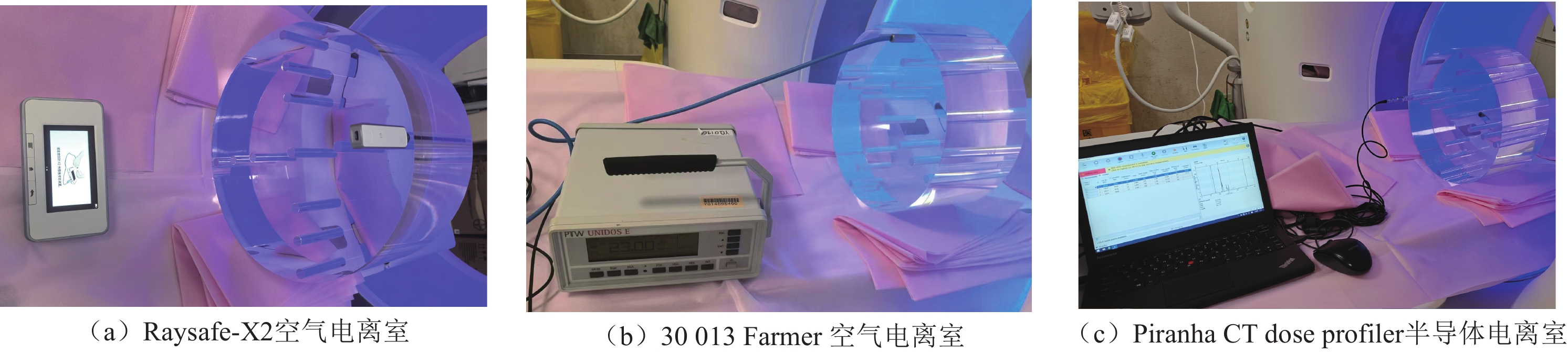
Objective: The point dose and volume CT dosimetry index (CTDIvol) at different X-ray energies were measured using the Raysafe-X2 air ionization chamber (referred to as “RX2”), the Piranha CT dose profiler semiconductor ionization chamber (referred to as “CDP”), and the
| [1] |
SMITH B R, WANG Y, CHU P, et al. International variation in radiation dose for computed tomography examinations: prospective cohort study[J]. BioMed Central, 2019, 364 k4931. DOI: 10.1136/bmj.k4931.
|
| [2] |
MCALLISTER K, LORIMORE S, WRIGHT E, et al. In vivo interactions between ionizing radiation, inflammation and chemical carcinogens identified by increased DNA damage responses[J]. Radiation Research, 2012, 177(5): 584-593. DOI: 10.1667/rr2690.1.
|
| [3] |
POON R, BADAWY M K. Radiation dose and risk to the lens of the eye during CT examinations of the brain[J]. Journal of Medical Imaging and Radiation Oncology, 2019, 63(6): 786-794. DOI: 10.1111/1754-9485.12950.
|
| [4] |
BOSCH M, THIERRY I, HARBRON R, et al. Risk of hematological malignancies from CT radiation exposure in children, adolescents and young adults[J]. Nature Medicine, 2023, 29(12): 3111-3119. DOI: 10.1038/s41591-023-02620-0.
|
| [5] |
HAUPTMANN M, DANIELS RD, CARDIS E, et al. Epidemiological Studies of Low-Dose Ionizing Radiation and Cancer: Summary Bias Assessment and Meta-Analysis[J]. J Natl Cancer Inst Monogr, 2020(56): 188-200. DOI: 10.1093/jncimonographs/lgac027.
|
| [6] |
王楚胭, 卓维海, 林鑫, 等. CT所致受检者个体化器官剂量的研究进展[J]. 中国辐射卫生, 2022, 31(6): 756-762. DOI: 10.13491/j.issn.1004-714X.2022.06.021.
WANG C Y, ZHUO W H, LIN X, et al. Research progress on individualized organ doses for patients caused by CT[J]. Chinese Journal of Radiation Health, 2022, 31(6): 756-762. DOI: 10.13491/j.issn.1004-714X.2022.06.021.
|
| [7] |
欧向明, 范瑶华. 新型诊断剂量仪的能量响应特性研究[J]. 中国医学装备, 2018, 15(7): 58-60. DOI: 10.3969/J.ISSN.1672-8270.2018.07.013.
OU X M, FAN Y H. Energy response characteristics of a new diagnostic dose meter[J]. China Medical Equipment, 2018, 15(7): 58-60. DOI: 10.3969/J.ISSN.1672-8270.2018.07.013.
|
| [8] |
卫生健康委员会. X 射线计算机体层摄影装置质量控制检测规范: WS/T 519-2019[S]. 2019.
|
| [9] |
郭洪涛, 刘勇, 袁淑华. CT剂量指数(CTDI)测量研究[J]. 中国测试技术, 2007, 33(4): 33-36,108. DOI: 10.3969/j.issn.1674-5124.2007.04.009.
GUO H T, LIU Y, YUAN S H. Research on CT dose index (CTDI) measurement[J]. China Testing Technology, 2007, 33(4): 33-36,108. DOI: 10.3969/j.issn.1674-5124.2007.04.009.
|
| [10] |
杨志国. 检定螺旋CT剂量指数(CTDI)应注意的问题[J]. 中国计量, 2015(2): 115-116.
YANG Z G. Issues to be Noted in the Calibration of Spiral CT Dose Index (CTDI)[J]. China Metrology, 2015(2): 115-116. (in Chinese).
|
| [11] |
徐少一, 李伟, 廖凯锋, 等. 硅半导体辐射探测仪表国产化研制及性能研究[J]. 核电子学与探测技术, 2024, 44(4): 608-614. DOI: 10.3969/j.issn.0258-0934.2024.04.003.
XU S Y, LI W, LIAO K F, et al. Research and Development of Domestic Silicon Semiconductor Radiation Detection Instruments and Their Performance[J]. Nuclear Electronics and Detection Technology, 2024, 44(4): 608-614. DOI: 10.3969/j.issn.0258-0934.2024.04.003.
|
| [12] |
郑海亮, 李兴东, 刘小丽, 等. CT-SD16探测器辐射剂量测量原理和参数设定[J]. 北京生物医学工程, 2012, 31(3): 278-282. DOI: 10.3969/j.issn.1002-3208.2012.03.12.
ZHENG H L, LI X D, LIU X L, et al. Radiation Dose Measurement Principles and Parameter Settings of CT-SD16 Detector[J]. Beijing Biomedical Engineering, 2012, 31(3): 278-282. DOI: 10.3969/j.issn.1002-3208.2012.03.12.
|
| [13] |
庄静文, 郑钧正, 白玫. CT剂量指数估算方法研究[J]. 中国医学装备, 2016, 13(7): 1-3,4. DOI: 10.3969/J.ISSN.1672-8270.2016.07.001.
ZHUANG J W, ZHENG J Z, BAI M. Research on Estimation Methods for CT Dose Index[J]. China Medical Equipment, 2016, 13(7): 1-3,4. DOI: 10.3969/J.ISSN.1672-8270.2016.07.001.
|
| [14] |
沈春花. 四种不同型号诊断X射线剂量仪的剂量探测技术概述[J]. 中国医学装备, 2005, 2(10): 33-34. DOI: 10.3969/j.issn.1672-8270.2005.10.013.
SHEN C H. Overview of Dose Detection Technologies for Four Different Models of Diagnostic X-ray Dose Meters[J]. China Medical Equipment, 2005, 2(10): 33-34. DOI: 10.3969/j.issn.1672-8270.2005.10.013.
|
| [15] |
陈坤锋, 罗雄峰, 项毅, 等. 140mm电离室检定宽射线束CT辐射剂量指数的不确定度评定[J]. 仪器仪表标准化与计量, 2022(6): 32-34. DOI: 10.3969/j.issn.1672-5611.2022.06.011.
CHEN K F, LUO X F, XIANG Y , et al. Uncertainty Assessment of 140mm Ionization Chamber for Calibration of Wide X-ray Beam CT Radiation Dose Index [J]. Instrumentation and Metrology, 2022(6): 32-34. DOI: 10.3969/j.issn.1672-5611.2022.06.011.(in Chinese).
|
| [16] |
郑海亮, 李兴东, 张鹏, 等. 多排螺旋CT五个剂量测量点位的统计学差异[J]. CT理论与应用研究, 2015, 24(3): 337-343. DOI: 10.15953/j.1004-4140.2015.24.03.02.
ZHENG H L, LI X D, ZHANG P, et al. Statistical Differences of Five Dose Measurement Points in Multislice Spiral CT[J]. CT Theory and Applications Research, 2015, 24(3): 337-343. DOI: 10.15953/j.1004-4140.2015.24.03.02.
|
Supported by: Beijing Renhe Information Technology Co. Ltd 


 DownLoad: