ISSN 1004-4140
CN 11-3017/P
| Citation: |
WHANG C X, CHEN P, PAN J X, et al. Research on material decomposition of dual-energy CT image based on iterative residual network[J]. CT Theory and Applications, 2022, 31(1): 47-54. DOI: 10.15953/j.1004-4140.2022.31.01.05. (in Chinese).

|
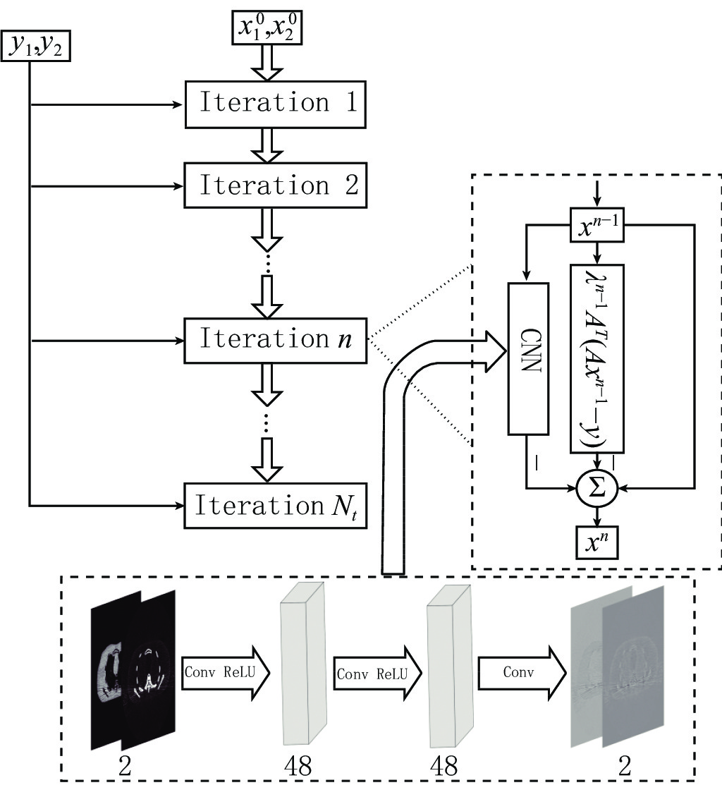
Dual energy computed tomography (DECT) plays an important role in the application of advanced imaging due to its material decomposition capability. Image domain decomposition can directly invert CT images through by linear matrix, but the decomposed material images will be seriously affected by noise and artifacts. Although various regularization methods have been proposed to solve this problem, they still face two challenges: tedious parameter adjustment and the loss of image details resulted from over-smoothing. Therefore, in this paper we proposes a dual energy CT image material decomposition algorithm based on iterative residual network. Direct inversion is used as the initial base image, and a stacking two-channel convolutional neural network is used to replace the regularization items in the iterative decomposition model to form a deep iterative decomposition network. This method can realize material decomposition and noise suppression simultaneously. Experimental results show that the iterative residual network proposed in this paper is superior to other comparison methods and can effectively suppress noise and artifacts while maintaining the edge details of the base image.
| [1] |
JOHNSON T R, KRAU B, SEDLMAIR M, et al. Material differentiation by dual energy CT: Initial experience[J]. European Radiology, 2007, 17(6): 1510-1517.
|
| [2] |
王文杰, 陈平, 潘晋孝, 等. 基于参考组分的双能CT成像方法[J]. CT理论与应用研究, 2021,30(1): 61−69. DOI: 10.15953/j.1004-4140.2021.30.01.06.
WANG W J, CHEN P, PAN J X, et al. Dual-energy CT imaging method based on reference components[J]. CT Theory and Applications, 2021, 30(1): 61−69. DOI: 10.15953/j.1004-4140.2021.30.01.06. (in Chinese).
|
| [3] |
王李磊. 双能CT 图像重建算法研究[D]. 郑州: 解放军信息工程大学, 2016.
WANG L L. Research on dual energy CT image reconstruction algorithm[D]. Zhengzhou: PLA University of information engineering, 2016. (in Chinese).
|
| [4] |
XUE Y, RUAN R S, HU X H, et al. Statistical image-domain multimaterial decomposition for dual-energy CT[J]. Medical Physics, 2017, 44(3): 886−901. doi: 10.1002/mp.12096
|
| [5] |
XUE D, NIU T Y, ZHU L. Combined iterative reconstruction and image-domain decomposition for dual energy CT using total-variation regularization[J]. Medical Physics, 2014, 41(5): 051909. DOI: 10.1118/1.4870375.
|
| [6] |
DING Q Q, NIU T Y, ZHANG X Q, et al. Image-domain multi-material decomposition for dual-energy CT based on prior information of material images[J]. Medical Physics, 2018: 45. DOI: 10.1002/mp.13001.
|
| [7] |
LI Z, RAVISHANKAR S, YONG L, et al. Image-domain material decomposition using data-driven sparsity models for dual-energy CT[C]//2018 IEEE 15th International Symposium on Biomedical Imaging (ISBI 2018). IEEE, 2018.
|
| [8] |
LI Z P, RAVISHANKAR S, LONG Y, et al. DECT-MULTRA: Dual-energy CT image decomposition with learned mixed material models and efficient clustering[J]. IEEE Transactions on Medical Imaging, 2020, 39(4): 1223−1234. DOI: 10.1109/TMI.2019.2946177.
|
| [9] |
ZHANG J L, GU Y B, TANG H, et al. Compressed sensing MR image reconstruction via a deep frequency-division network[J]. Neurocomputing, 2020, 384: 346−355. doi: 10.1016/j.neucom.2019.12.011
|
| [10] |
CHEN H, ZHANG Y, KALRA M K, et al. Low-dose CT with a residual encoder-decoder convolutional neural network (RED-CNN)[J]. IEEE Transactions on Medical Imaging, 2017, 36(99): 2524−2535.
|
| [11] |
SU T, SUN X, ZHANG Y, et al. DIRECT-net: A unified mutual-domain material decomposition network for quantitative dual-energy CT imaging[J]. 2020.
|
| [12] |
CHEN H, ZHANG Y, CHEN Y, et al. LEARN: Learned experts' assessment-based reconstruction network for sparse-data CT[J]. IEEE Transactions on Medical Imaging, 2018, (99): 1−1.
|
| [13] |
YANG Y, SUN J, LI H, et al. ADMM-CSNet: A deep learning approach for image compressive sensing[J]. IEEE Transactions on Pattern Analysis and Machine Intelligence, 2020: 521−538.
|
| [14] |
ADLER J, OKTEM O. Learned primal-dual reconstruction[J]. IEEE Transactions on Medical Imaging, 2017: 1322−1332.
|
| [15] |
XU Y F, YAN B, ZHANG J F, et al. Image decomposition algorithm for dual-energy computed tomography via fully convolutional network[J]. Computational & Mathematical Methods in Medicine, 2018: 2527516. DOI: 10.1155/2018/2527516.
|
Supported by: Beijing Renhe Information Technology Co. Ltd 


 DownLoad: