ISSN 1004-4140
CN 11-3017/P
| Citation: |
SUN Y, LI L, LIU X Y, et al. Imaging Features of Early COVID-19 on Chest Thin-slice Non-Enhanced CT[J]. CT Theory and Applications, 2023, 32(1): 131-138. DOI: 10.15953/j.ctta.2023.006. (in Chinese).

|
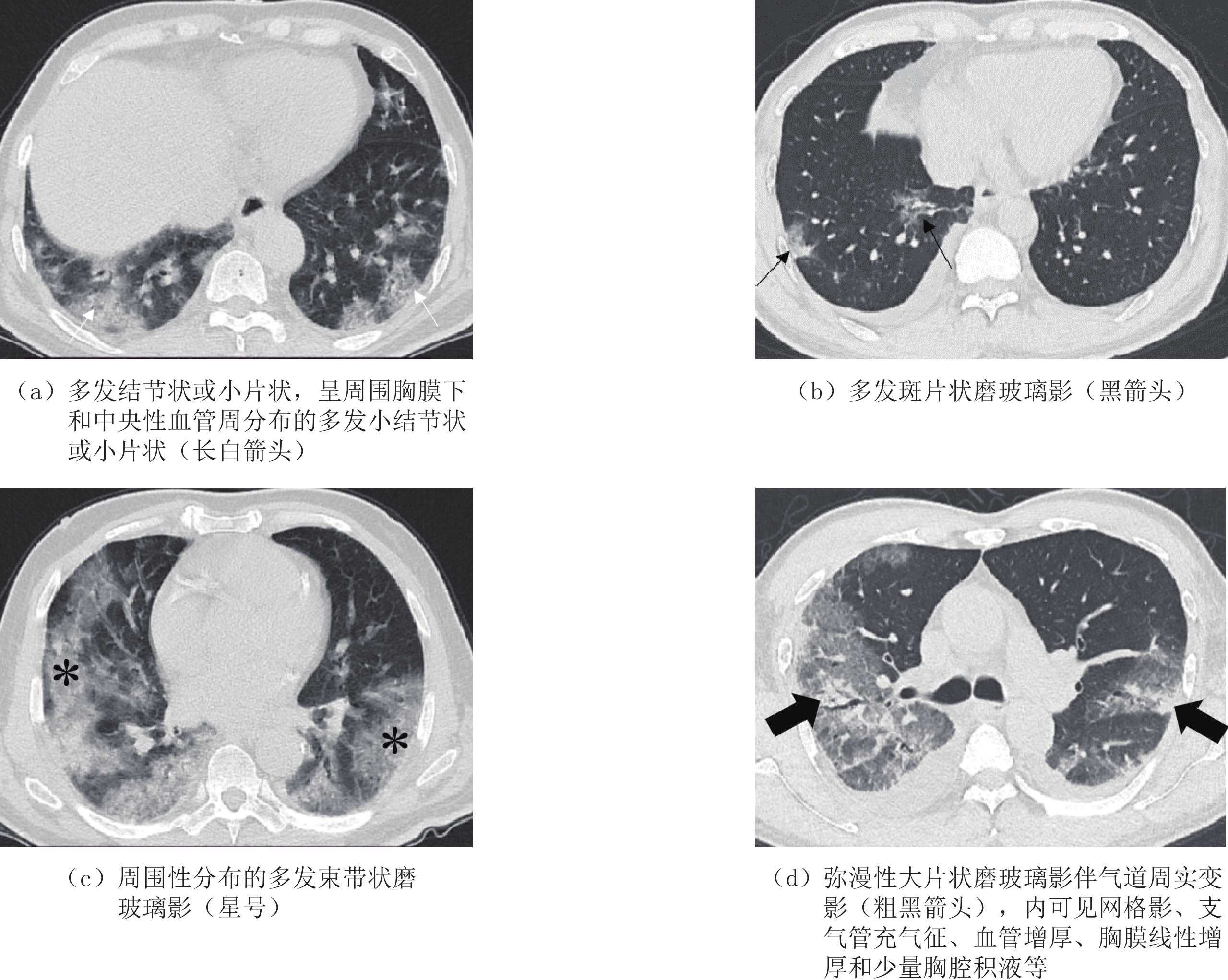
Objective: To explore the characteristics findings of early COVID-19 on chest thin-slice non-enhanced CT. Materials and methods: From November to December 2022, data from 153 patients with COVID-19 with positive chest CT findings confirmed by the Department of Infectious Diseases of our hospital were collected. All patients had relatively complete clinical data and underwent chest CT 1-14 days after the onset. The patients were divided into two groups according to their age (≤60 years old and >60 years old) and the time interval between the onset and CT examination (≤7 days old and >7 days old), and the differences in CT features between the two groups were compared. Result: Among 153 patients with COVID-19, the lung (100%), blood vessels (93.5%), airways (73.4%), and pleura (70.1%) were involved. The comparison between the age groups showed statistically significant differences in the number, location, size, volume, and band shadow of the lesions. There were statistically significant differences in the shape, density, organization, fibrosis, and pleural involvement of the lesions between the patients in the time interval between the onset and CT examination. Conclusion: Chest thin-slice CT can clarify the location and scope of early COVID-19. Some characteristics findings can help for accurate diagnosis and differentiation, such as the diversity of morphology, intrapleural and perivascular distribution, interstitial lesions with alveoli as a unit, early mixed ground-glass shadow often accompanied by obvious organization and fibrosis, localized thickening of the pleura, and few pleural effusions.
| [1] |
XIE J, WANG Q, XU Y, et al. Clinical characteristics, laboratory abnormalities and CT findings of COVID-19 patients and risk factors of severe disease: A systematic review and meta-analysis[J]. Annals of Palliative Medicine, 2021, 10(2): 1928-1949.
|
| [2] |
LIU Y, ZHOU X, LIU X, et al. Systematic review and meta-analysis of the CT imaging characteristics of infectious pneumonia[J]. Annals of Palliative Medicine, 2021, 10(10): 10414−10424. doi: 10.21037/apm-21-2101
|
| [3] |
Tang Y, Liao H, Wu Q, et al. Chest CT imaging characteristics and their evolution of 48 patients with COVID-19 in Hengyang, China[J]. American Journal of Translational Research, 2021, 13(9): 9983−9992.
|
| [4] |
PAKDEMIRLI E, MANDALIA U, MONIB S. Characteristics of Chest CT images in patients with COVID-19 Pneumonia in London, UK[J]. Cureus, 2020, 12(9): e10289.
|
| [5] |
MARCHIORI E, NOBRE L F, HOCHHEGGER B, et al. CT characteristics of COVID-19: Reversed halo sign or target sign?[J]. Diagnostic and Interventional Radiology, 2021, 27(2): 306−307. doi: 10.5152/dir.2020.20734
|
| [6] |
HUANG C, WANG Y, LI X, et al. Clinical features of patients infected with 2019 novel coronavirus in Wuhan, China[J]. Lancet, 2020, 395(10223): 497−506. doi: 10.1016/S0140-6736(20)30183-5
|
| [7] |
LIU H, LUO S, LI H, et al. Clinical characteristics and longitudinal chest CT features of healthcare workers hospitalized with coronavirus disease 2019 (COVID-19)[J]. International Journal of Medical Sciences, 2020, 17(17): 2644−2652. doi: 10.7150/ijms.48696
|
| [8] |
JAFARI R, MAGHSOUDI H, SABURI A. A unique feature of COVID-19 infection in chest CT; "Pulmonary Target" appearance[J]. Academic Radiology, 2021, 28(1): 146−147. doi: 10.1016/j.acra.2020.11.004
|
| [9] |
WU R, GUAN W, GAO Z, et al. The arch bridge sign: A newly described CT feature of the coronavirus disease-19 (COVID-19) pneumonia[J]. Quantitative Imaging in Medicine and Surgery, 2020, 10(7): 1551−1558. doi: 10.21037/qims-20-688
|
| [10] |
YOON S H, LEE J H, KIM B N. Chest CT findings in hospitalized patients with SARS-CoV-2: Delta versus Omicron Variants[J]. Radiology, 2023, 306(1): 252−260. doi: 10.1148/radiol.220676
|
| [11] |
黄益龙, 张振光, 李翔, 等. CT影像组学联合征象鉴别新型冠状病毒肺炎与其他病毒性肺炎的价值[J]. 中华放射学杂志, 2022,56(1): 36−42. doi: 10.3760/cma.j.cn112149-20201220-01318
HUANG Y L, ZHANG Z G, LI X H, et al. The value of CT signs combined with radiomics in the differentiation of COVID-19 from other viral pneumonias[J]. Chinese Journal of Radiology, 2022, 56(1): 36−42. (in Chinese). doi: 10.3760/cma.j.cn112149-20201220-01318
|
| [12] |
赵小二, 邓克学, 王朋. 不同阶段新型冠状病毒肺炎的CT影像演变分析[J]. 实用放射学杂志, 2021,37(8): 1254−1257. doi: 10.3969/j.issn.1002-1671.2021.08.008
ZHAO X E, DENG K X, WANG P. Analysis of the CT manifestations changes of COVID-19 at different stages[J]. Journal of Practical Radiology, 2021, 37(8): 1254−1257. (in Chinese). doi: 10.3969/j.issn.1002-1671.2021.08.008
|
| [13] |
吴杰, 肖安岭, 顾金凤. 多层螺旋CT对新型冠状病毒肺炎的临床诊断价值[J]. 实用放射学杂志, 2021,37(5): 746−748. doi: 10.3969/j.issn.1002-1671.2021.05.013
WU J, XIA0 A L, GU J F. Clinical value of MSCT in the diagnosis of COVID-19[J]. Journal of Practical Radiology, 2021, 37(5): 746−748. (in Chinese). doi: 10.3969/j.issn.1002-1671.2021.05.013
|
| [14] |
余成成, 杨彦鸿, 胡天丽, 等. 新型冠状病毒B.1.617.2变异株感染者高分辨率CT与临床特点[J]. 中华放射学杂志, 2021,55(10): 1054−1058.
YU C C, YANG Y H, HU T L, et al. High resolution CT findings and clinical features of the novel coronavirus B.1.617.2 variant[J]. Journal of Practical Radiology, 2021, 55(10): 1054−1058. (in Chinese).
|
| [15] |
李声鸿, 曾献军, 鄢海蓝, 等. 新型冠状病毒肺炎薄层CT评价[J]. 实用放射学杂志, 2021,37(7): 1074−1076,1130. doi: 10.3969/j.issn.1002-1671.2021.07.007
LI S H, ZENG X J, YAN H L, et al. Evaluation of COVID-19 with thin-sclice CT[J]. Journal of Practical Radiology, 2021, 37(7): 1074−1076,1130. (in Chinese). doi: 10.3969/j.issn.1002-1671.2021.07.007
|
| [16] |
许玉环, 吕晓艳, 张见增, 等. 新型冠状病毒肺炎不同临床分型的CT特征[J]. 中国医学影像学杂志, 2020,28(12): 887−890, 895. doi: 10.3969/j.issn.1005-5185.2020.12.002
XU Y H, LV X Y, ZHANG J Z, et al. CT features of different clinical types of CoVID-19[J]. Chinese Journal of Medical Imaging, 2020, 28(12): 887−890, 895. (in Chinese). doi: 10.3969/j.issn.1005-5185.2020.12.002
|
| [17] |
余鎏, 肖易, 赵泉. 新型冠状病毒肺炎的临床表现及胸部高分辨率CT影像学表现分析[J]. 实用放射学杂志, 2021,37(7): 1081−1085. doi: 10.3969/j.issn.1002-1671.2021.07.009
YU L, XIAO Y, ZHAO Q. The clinical manifestations and chest high resolution CT findings of patients with COVID-19[J]. Journal of Practical Radiology, 2021, 37(7): 1081−1085. (in Chinese). doi: 10.3969/j.issn.1002-1671.2021.07.009
|
| [18] |
蒋玮丽, 龙斌, 柏玉涵, 等. 新型冠状病毒肺炎的胸部CT特征[J]. 中国医学影像学杂志, 2020,28(11): 817−819, 824. doi: 10.3969/j.issn.1005-5185.2020.11.005
JIANG W L, LONG B, BAI Y H, et al. Chest CT features of COVID-19[J]. Chinese Journal of Medical Imaging, 2020, 28(11): 817−819, 824. (in Chinese). doi: 10.3969/j.issn.1005-5185.2020.11.005
|
| [19] |
纪丙军, 齐庆梅, 王聪, 等. 新型冠状病毒肺炎与其他社区获得性肺炎不同病期的CT表现及动态分析[J]. 实用放射学杂志, 2021,37(8): 1266−1270. doi: 10.3969/j.issn.1002-1671.2021.08.011
JI B J, QI Q M, WANG C, et al. CT manifestations and dynamic change of different stages of COVID-19 and other community acquired pneumonia[J]. Journal of Practical Radiology, 2021, 37(8): 1266−1270. (in Chinese). doi: 10.3969/j.issn.1002-1671.2021.08.011
|
| [20] |
张庆, 熊浩, 彭婕, 等. 胸部CT对新型冠状病毒肺炎的诊断价值[J]. 中国医学影像学杂志, 2020,28(12): 896−898. doi: 10.3969/j.issn.1005-5185.2020.12.004
ZHANG Q, XIONG H, PENG J, et al. Diagnostic value of chest CT for COVID-19[J]. Chinese Journal of Medical Imaging, 2020, 28(12): 896−898. (in Chinese). doi: 10.3969/j.issn.1005-5185.2020.12.004
|
| [1] | KANG Zhaoting, OUYANG Xuehui, CHAI Jun. Differential Diagnosis of COVID-19 and Community-acquired Pneumonia Using Different Machine Learning Methods[J]. CT Theory and Applications, 2023, 32(5): 685-694. DOI: 10.15953/j.ctta.2023.079 |
| [2] | LIU Xiaoyan, BAO Zhongying, DUAN Shuhong, ZHANG Jie, ZHANG Mingxia, SUN Ying, LI Ling, WANG Rengui. Clinical Characteristics and Imaging Features of COVID-19 at Initial Diagnosis in Fever Clinic[J]. CT Theory and Applications, 2023, 32(5): 636-644. DOI: 10.15953/j.ctta.2023.149 |
| [3] | ZHANG Kai, CHAI Jun, LIU Rui, ZHAO Jianhua, WANG Jingchen. Quantitative Analysis of Computed Tomography Features of Different COVID-19 Infection Virus Variants Using Artificial Intelligence[J]. CT Theory and Applications, 2023, 32(5): 595-602. DOI: 10.15953/j.ctta.2023.043 |
| [4] | WANG Rengui. Clinical, Pathological and Imaging Manifestations of Novel Coronavirus Infection[J]. CT Theory and Applications, 2023, 32(3): 297-302. DOI: 10.15953/j.ctta.2023.116 |
| [5] | ZHANG Fengling, ZHAO Li, LIU Jiabao. CT Features of Pulmonary Infection in Elderly Patients with Type 2 Diabetes Mellitus[J]. CT Theory and Applications, 2021, 30(5): 583-590. DOI: 10.15953/j.1004-4140.2021.30.05.06 |
| [6] | SHEN Jing, YU Jing, YAN Yingnan, SANG Yarong, JU Ronghui, PAN Long, LI Guize, LI Xin, WU Jianlin. Chest CT Features of COVID-19 and Its Evolution[J]. CT Theory and Applications, 2021, 30(2): 199-207. DOI: 10.15953/j.1004-4140.2021.30.02.07 |
| [7] | ZHANG Hecheng, CHU Yan, LIU Jing, LI Xiaozhen, ZHAO Tianzuo. The Clinical Features and CT Manifestations of the Novel Coronavirus Pneumonia COVID-19[J]. CT Theory and Applications, 2020, 29(5): 559-565. DOI: 10.15953/j.1004-4140.2020.29.05.06 |
| [8] | YAO Yonggang, DU Jingbo, LIAO Jianyong, GOU Zhenheng, FU Shunbin, JIN Erhu. Study of Chest CT Features of COVID-19[J]. CT Theory and Applications, 2020, 29(2): 169-176. DOI: 10.15953/j.1004-4140.2020.29.02.07 |
| [9] | CHEN Yuehua, ZHANG Tao. Clinical Features and CT Imaging Findings of Patients with Corona Virus Disease-19[J]. CT Theory and Applications, 2020, 29(2): 155-162. DOI: 10.15953/j.1004-4140.2020.29.02.05 |
| [10] | WU Yan-chun, LIU Yao, YAN Zhi-han. Exploration of CT Imaging Features of Chronic Severe Hepatitis Complicated with Invasive Pulmonary Fungal Infection[J]. CT Theory and Applications, 2017, 26(4): 473-479. DOI: 10.15953/j.1004-4140.2017.26.04.09 |
Supported by: Beijing Renhe Information Technology Co. Ltd 


 DownLoad: