ISSN 1004-4140
CN 11-3017/P
| Citation: |
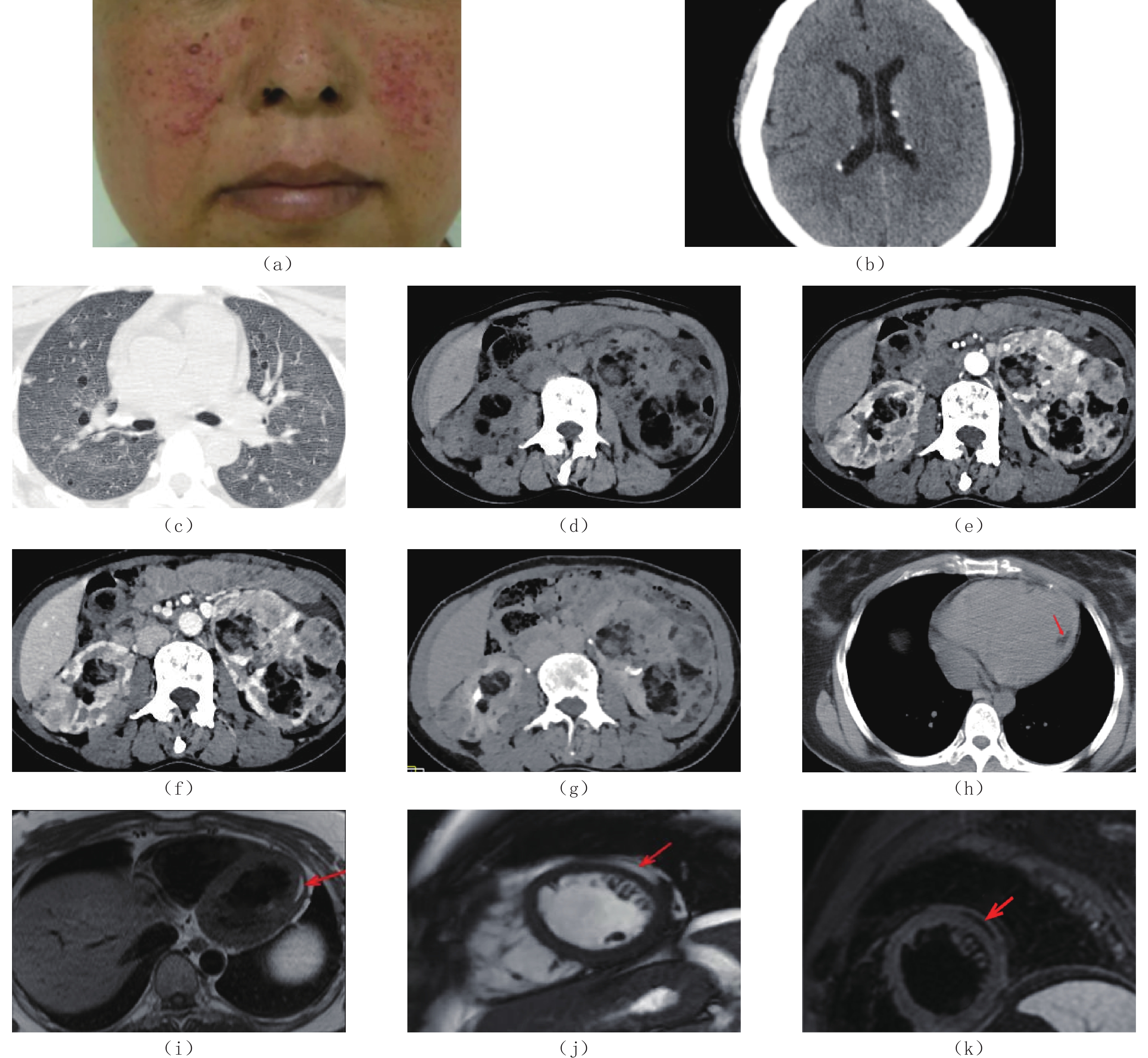
LI Y, XIE K. Diagnosis of Tuberous Sclerosis Complex with Multiple Organ Involvement using Computer Tomography: A Clinical Case Analysis[J]. CT Theory and Applications, 2024, 33(3): 359-364. DOI: 10.15953/j.ctta.2023.112. (in Chinese).

|
Tuberous sclerosis complex (TSC) is an autosomal dominant genetic disease caused by mutations in the tumor suppressor genes TSC1 and/or TSC2. Its clinical manifestations include recurrent seizures, intellectual disability, and facial angiofibromas. Herein, we report the case of a patient with TSC who had recurrent seizures since childhood, shark skin nevus on the right occipital region, and multiple purple-red nodules on both cheeks. Computer tomography (CT) revealed multiple small calcifications around the lateral ventricles, pulmonary lymphangioleiomyomatosis, multiple renal angiomyolipomas, and multiple lipomatous lesions on the left ventricular wall. Through this case report, we aimed to improve the understanding of TSC.
| [1] |
NORTHRUP H, KRUEGER D A. Tuberous sclerosis complex diagnostic criteria update: Recommendations of the 2012 international tuberous sclerosis complex consensus conference[J]. Pediatric Neurology, 2013, 49(4): 243−254.
|
| [2] |
PORTOCARRERO L K L, QUENTAL K N, SAMORANO L P, et al. Tuberous sclerosis complex: Review based on new diagnostic criteria[J]. Anais Brasileiros de Dermatologia, 2018, 93(3): 323−331.
|
| [3] |
Von RANKE F M, FARIA I M, ZANETTI G, et al. Imaging of tuberous sclerosis complex: A pictorial review[J]. Radiologia Brasileira, 2017, 50(1): 48−54. DOI: 10.1590/0100-3984.2016.0020.
|
| [4] |
RODRIGUES D A, GOMES C M, COSTA I M. Tuberous sclerosis complex[J]. Anais Brasileiros de Dermatologia, 2012, 87(2): 184−196. DOI: 10.1590/S0365-05962012000200001.
|
| [5] |
CURATOLO P, MOAVERO R, de VRIES P J. Neurological and neuropsychiatric aspects of tuberous sclerosis complex[J]. Lancet Neurology, 2015, 14(7): 733−745. DOI: 10.1016/S1474-4422(15)00069-1.
|
| [6] |
RITTER D M, FESSLER B K, EBRAHIMI-FAKHARI D, et al. Prevalence of thoracoabdominal imaging findings in tuberous sclerosis complex[J]. Orphanet Journal of Rare Diseases, 2022, 17(1): 124−134.
|
| [7] |
MIZUGUCHI M, OHSAWA M, KASHII H, et al. Brain symptoms of tuberous sclerosis complex: Pathogenesis and treatment[J]. International Journal of Molecular Sciences, 2021, 22(13): 6677−6692.
|
| [8] |
LIN Y, WU W, GAO L, et al. Multimodality imaging of benign primary cardiac tumor[J]. Diagnostics (Basel), 2022, 12(10): 2543−2562.
|
| [9] |
HOEY E T, MANKAD K, PUPPALA S, et al. MRI and CT appearances of cardiac tumours in adults[J]. Clinical Radiology, 2009, 64(12): 1214−1230. DOI: 10.1016/j.crad.2009.09.002.
|
| [10] |
ALSHOABI S A, HAMID A M, ALHAZMI F H, et al. Diagnostic features of tuberous sclerosis complex: Case report and literature review[J]. Quantitative Imaging in Medicine and Surgery, 2022, 12(1): 846−861.
|
| [11] |
王乐, 秦娜, 郝美美, 等. 中国结节性硬化症患者的临床特点及诊断现状分析[J]. 现代生物医学进展, 2019, 19(8): 1567−1572.
WANG L, QIN N, HAO M M, et al. Clinical features and diagnosis of patients with tuberous sclerosis complex, a current evaluation in China[J]. Progress in Modern Biomedicine, 2019, 19(8): 1567−1572. (in Chinese).
|
| [12] |
ILYAS M, QUEZADA J, OPFER E K. Lipomatous infiltration in tuberous sclerosis complex: A case series and literature review[J]. Child Neurology Open, 2021, 20(8): 48−65.
|
| [13] |
LUO C, YE W R, SHI W, et al. Perfect match: mTOR inhibitors and tuberous sclerosis complex[J]. Orphanet Journal of Rare Diseases, 2022, 17(1): 106−109. DOI: 10.1186/s13023-022-02266-0.
|
| [14] |
PENG J H, TU H P, HONG C H. A population-based study to estimate survival and standardized mortality of tuberous sclerosis complex (TSC) in Taiwan[J]. Orphanet Journal of Rare Diseases, 2021, 16(1): 335−348.
|
| [15] |
李林, 赵建设. MRI是儿童先天性TORCH综合征治疗的首选影像学检查方式[J]. 分子影像学杂志, 2019, 42(4): 430−433.
LI L, ZHAO J S. Application and clinical analysis of MRI in the TORCH of children[J]. Journal of Molecular Imaging, 2019, 42(4): 430−433. (in Chinese).
|
| [16] |
CIPRIANI A, MATTESI G, BARIANI R, et al. Cardiac magnetic resonance imaging of arrhythmogenic cardiomyopathy: Evolving diagnostic perspectives[J]. European Radiology, 2023, 33(1): 270−282.
|
| [1] | REN Yue, ZHANG Yongxian, LIU Dandan, MA Wentao, GUO Senlin. Application of Bismuth Shielding and Organ Dose Modulation Techniques in Brain CT Scanning with Different Scanning Baselines[J]. CT Theory and Applications, 2025, 34(3): 345-350. DOI: 10.15953/j.ctta.2024.318 |
| [2] | Yin Wei, He quanyu, Yin Hongxia, Qin Xiangyu, Wu Kewei, Zhong Zhaohui. Exploration of Organ Dose Modulation in Male Pelvic CT Scanning[J]. CT Theory and Applications. DOI: 10.15953/j.ctta.2024.307 |
| [3] | ZHANG Yongxian, LIU Dandan. Combined Application of Bismuth Shielding and Organ Dose-Modulation Techniques in Lung CT Scanning[J]. CT Theory and Applications. DOI: 10.15953/j.ctta.2025.092 |
| [4] | SUI Zhao, YAN Xiaohu, LI Ying. Analysis on Image Features of Tuberculous Sclerosis in Different Parts of the Skull in CT Scan[J]. CT Theory and Applications, 2020, 29(6): 695-701. DOI: 10.15953/j.1004-4140.2020.29.06.07 |
| [5] | HU Yin-song, HOU Zhi-xiong, DENG Jia-xiu, LI Guo-qiang, LAN Hua, HONG Jian-bin. CT Diagnosis of Neonatal Hypoxic-Ischemic Encephalopathy[J]. CT Theory and Applications, 2011, 20(4): 531-535. |
| [6] | XU Guan-zhen, FU Chuan-ming, JIANG Lin. CT Diagnosis of the External Hydrocephalus[J]. CT Theory and Applications, 2011, 20(3): 397-401. |
| [7] | WAN Xia, BI Chun-long. Radiological Image Manifestations of Tuberous Sclerosis[J]. CT Theory and Applications, 2006, 15(3): 35-39. |
| [8] | Bi Chunlong, Gao Liyuan, Wan Xia. CT Diagnosis of Cerebral Tuberous Sclerosis in Infants and Children[J]. CT Theory and Applications, 2001, 10(1): 28-29. |
| [9] | Gao Yan, Xu Junchao. CT Diagnosis of Acute Cervical Trauma[J]. CT Theory and Applications, 2000, 9(4): 35-39. |
| [10] | Gui Chengli, Luan Kedong, Mo Yajun. The Analysis of CT and Clinical Data of External Hydrocephalus[J]. CT Theory and Applications, 2000, 9(3): 23-25. |
Supported by: Beijing Renhe Information Technology Co. Ltd 


 DownLoad: