ISSN 1004-4140
CN 11-3017/P
| Citation: |
LI J J, LIU Y D, LI X T, et al. Correlations of Hip Muscle Composition with Age and Body Mass Index in Middle-aged and Elderly Patients with Hip Fracture[J]. CT Theory and Applications, 2024, 33(5): 619-625. DOI: 10.15953/j.ctta.2023.143. (in Chinese).

|
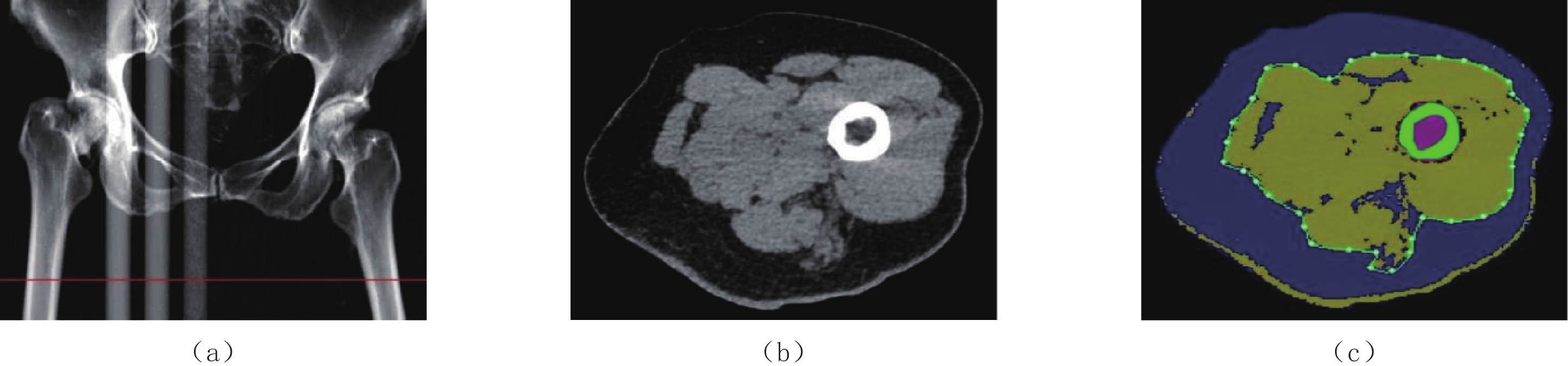
Objective: To investigate the correlations of hip muscle composition with age and body mass index (BMI) in middle-aged and elderly patients with hip fracture. Methods: In total of 175 patients aged 51~95 years with low-energy hip fracture who underwent hip computed tomography scans were divided into <80 and ≥80 year-old groups. Quantitative computed tomography (QCT) was performed to obtain the cross-sectional muscle area (CSMA), total adipose area (TAA), intra-muscular adipose area (IMAA), subcutaneous adipose area (SAA) and muscle fat infiltration (MFI) of the hip. An independent t-test was used to compare the differences between gender and groups, and correlation analysis was used to determine the relationships between hip muscle fat area and age and BMI. Results: The hip TAA, SAA and MFI of men were significantly lower than those of women (t=−2.356, −2.550, −3.090), and CSMA was significantly higher than that of women. The CSMA in the <80 year-old age group was higher than that of the ≥80 year-old age group, and the MFI was lower than that found in the ≥80 years age old group. After adjusting for BMI, the age of men and women positively correlated with IMAA and MFI (r=0.445 and 0.612 for men, r=0.202 and 0.390 for women), and negatively correlated with CSMA (r=−0.673 for men, r=−0.428 for women). After adjusting for age, the BMI of men positively correlated with TAA, IMAA, SAA, and CSMA (r=0.430, 0.491, 0.389, 0.623), whereas the BMI of women positively correlated with TAA, IMAA, SAA, CSMA, and MFI (r=0.510, 0.389, 0.478, 0.295, 0.296). Conclusion: In middle-aged and elderly patients with hip fragility fractures, men had more muscle and less total fat than those in women, but similar intermuscular fat to that of women. Hip muscle mass decreased and intermuscular fat increased with age. Generally, higher BMI, correlated with more muscle and fat. However, MFI in men was not associated with BMI.
| [1] |
SVEJME O, AHLBORG H G, NILSSON J A, et al. Low BMD is an independent predictor of fracture and early menopause of mortality in post-menopausal women: A 34-year prospective study[J]. Maturitas, 2013, 74(4): 341−345. DOI: 10.1016/j.maturitas.2013.01.002.
|
| [2] |
王玲,杨明辉,苏永彬,等. 老年女性髋部脆性骨折骨密度特点:病例对照研究[J]. 中华骨质疏松和骨矿盐疾病杂志, 2022, 15(1): 12−18. DOI: 10.3969/j.issn.1674-2591.2022.01.003.
WANG L, YANG M H, SU Y B, et al. Assessments of bone mineral density for elderly frailty hip fracture women with large sample size: A case control study[J]. Chinese Journal of Osteoporosis and Bone Mineral Research, 2022, 15(1): 12−18. DOI: 10.3969/j.issn.1674-2591.2022.01.003. (in Chinese).
|
| [3] |
中华医学会骨质疏松和骨矿盐疾病分会. 原发性骨质疏松症诊疗指南(2022)[J]. 中国全科医学,2023,26(14): 1671−1691.
Chinese Society of Osteoporosis and Bone Mineral Research. Guidelines for the diagnosis and treatment of primary osteoporosis(2022)[J]. Chinese General Practice, 2023, 26(14): 1671−1691. (in Chinese).
|
| [4] |
MALKOV S, CAWTHON P M, PETERS K W, et al. Hip fractures risk in older men and women associated with DXA-Derived measures of thigh subcutaneous fat thickness, cross-sectional muscle area, and muscle density[J]. Journal of Bone and Mineral Research, 2015, 30(8): 1414−1421. DOI: 10.1002/jbmr.2469.
|
| [5] |
HAHN M H, WON Y Y. Bone mineral density and fatty degeneration of thigh muscles measured by computed tomography in hip fracture patients[J]. Journal of Bone Metabolism, 2016, 23(4): 215−221. DOI: 10.11005/jbm.2016.23.4.215.
|
| [6] |
EDWARDS M H, DENNISON E M, AIHIE SAYER A, et al. Osteoporosis and sarcopenia in older age[J]. Bone, 2015, 80(11): 126−130.
|
| [7] |
HE H, LIU Y, TIAN Q, et al. Relationship of sarcopenia and body composition with osteoporosis[J]. Osteoporosis International, 2016, 27(2): 473−482. DOI: 10.1007/s00198-015-3241-8.
|
| [8] |
CHENG X, ZHANG Y, WANG C, et al. The optimal anatomic site for a single slice to estimate the total volume of visceral adipose tissue by using the quantitative computed tomography (QCT) in Chinese population[J]. European Journal of Clinical Nutrition, 2018, 72(11): 1567−1575. DOI: 10.1038/s41430-018-0122-1.
|
| [9] |
SHEN W, PUNYANITYA M, WANG Z, et al. Total body skeletal muscle and adipose tissue volumes: Estimation from a single abdominal cross-sectional image[J]. Journal of Applied Physiology, 2004, 97(6): 2333−2338. DOI: 10.1152/japplphysiol.00744.2004.
|
| [10] |
晏乘曦,王玲,姚丁华,等. CT定量测量髋部骨折患者髋部肌肉、脂肪面积及CT值的可重复性、可信度分析[J]. 山东医药,2018,58(16): 58−60. DOI: 10.3969/j.issn.1002-266X.2018.16.017.
|
| [11] |
LANG T F, SIGURDSSON S, KARLSDOTTIR G, et al. Age-related loss of proximal femoral strength in elderly men and women: The age Gene/environment susceptibility study--reykjavik[J]. Bone, 2012, 50(3): 743-748.
|
| [12] |
CRAWFORD R J, FILLI L, ELLIOTT J M, et al. Age- and level-dependence of fatty infiltration in lumbar paravertebral muscles of healthy volunteers[J]. American Journal of Neuroradiology, 2016, 37(4): 742−748. DOI: 10.3174/ajnr.A4596.
|
| [13] |
潘亚玲,陈彤彤,王晗琦,等. 定量CT分析年龄,腹部脂肪与骨密度的关系[J]. 中国医学影像学杂志,2020,28(4): 276−280. DOI: 10.3969/j.issn.1005-5185.2020.04.010.
PAN Y L, CHEN T T, WANG H Q, et al. Relationship among age, abdominal fat and bone mineral density by quantitative CT[J]. Chinese Journal of Medical Imaging, 2020, 28(4): 276−280. DOI: 10.3969/j.issn.1005-5185.2020.04.010. (in Chinese).
|
| [14] |
李新彤,钱占华,冯强强,等. 定量CT研究椎后肌群体质成分与年龄的关系及变化趋势[J]. 中华健康管理学杂志,2021,15(1): 44−48.
LI X T, QIAN Z H, FENG Q Q, et al. Relationship between posterior vertebral muscles composition and age by quantitative computed tomography[J]. Chinese Journal of Health Management, 2021, 15(1): 44−48. (in Chinese).
|
| [15] |
ILICH J Z, KELLY O J, INGLIS J E, et al. Interrelationship among muscle, fat, and bone: Connecting the dots on cellular, hormonal, and whole body levels[J]. Ageing Research Reviews, 2014, 15(5): 51−60.
|
| [16] |
WANG L, YIN L, ZHAO Y, et al. Muscle density discriminates hip fracture better than computed tomography X-ray absorptiometry hip areal bone mineral density[J]. Journal of Cachexia, Sarcopenia and Muscle, 2020, 11(6): 1799−1812. DOI: 10.1002/jcsm.12616.
|
| [17] |
VISSER M, KRITCHEVSKY S B, GOODPASTER B H, et al. Leg muscle mass and composition in relation to lower extremity performance in men and women aged 70 to 79: The health, aging and body composition study[J]. Journal of the American Geriatrics Society, 2002, 50(5): 897−904. DOI: 10.1046/j.1532-5415.2002.50217.x.
|
| [18] |
晏乘曦,王玲,姚丁华,等. 老年人躯干肌群与髋部肌群相关性研究[J]. 中国骨质疏松杂志,2018,24(12): 1586−1590. DOI: 10.3969/j.issn.1006-7108.2018.12.006.
YAN C X, WANG L, YAO D H, et al. Correlation between trunk muscle group and hip muscle[J]. Chinese Journal of Osteoporosis, 2018, 24(12): 1586−1590. DOI: 10.3969/j.issn.1006-7108.2018.12.006. (in Chinese).
|
| [19] |
Morley J E. Hormones and sarcopenia[J]. Current Pharmaceutical Design, 2017, 23(30): 4484−4492.
|
| [20] |
晁爱军,朱珊,胡玮,等. 不同性别体重指数与身体成份构成及脂肪分布的关系[J]. 中华骨质疏松和骨矿盐疾病杂志,2009,2(1): 24−27.
CHAO A J, ZHU S, HU W, et al. The relationship of body mass index with the body composing and fat distribution between men and women[J]. Chinese Journal of Osteoporosis and Bone Mineral Research, 2009, 2(1): 24−27. (in Chinese).
|
| [21] |
BOUTIN R D, YAO L, CANTER R J, et al. Sarcopenia: Current concepts and imaging implications[J]. American Journal of Roentgenology, 2015, 205(3): W255−266. DOI: 10.2214/AJR.15.14635.
|
| [22] |
MARCUS R L, ADDISON O, KIDDE J P, et al. Skeletal muscle fat infiltration: Impact of age, inactivity, and exercise[J]. Journal of Nutrition, Health, and Aging, 2010, 14(5): 362−366. DOI: 10.1007/s12603-010-0081-2.
|
| [23] |
ZENG Q, WANG L, DONG S, et al. CT-derived abdominal adiposity: Distributions and better predictive ability than BMI in a nationwide study of 59, 429 adults in China[J]. Metabolism, 2021, 115(2): 154456.
|
| [1] | LI Manman, FU Yigang, XIAO Yong, CHEN Wang, FENG Feng, XU Guodong. CT Radiomics Nomogram Prediction for Tumor Deposits and Prognosis in Colorectal Cancer[J]. CT Theory and Applications, 2025, 34(4): 694-702. DOI: 10.15953/j.ctta.2024.055 |
| [2] | XU Guodong, CHEN Wang, XIAO Yong, FU Yigang, WANG Manman, Li Manman. Prediction of postoperative disease-free survival in stage I–III colorectal cancer using a CT-based radiomics nomogram[J]. CT Theory and Applications. DOI: 10.15953/j.ctta.2025.043 |
| [3] | HU Zilin, WANG Shumei. Preliminary Study of CT Radiomics Analysis on Differentiating Exon 9/11 Mutations of c-kit Gene in Gastrointestinal Stromal Tumors[J]. CT Theory and Applications, 2022, 31(1): 73-79. DOI: 10.15953/j.1004-4140.2022.31.01.08 |
| [4] | XUE Ting, FENG Feng. Research Progress of Energy Spectrum CT of Colorectal Cancer[J]. CT Theory and Applications, 2020, 29(6): 751-758. DOI: 10.15953/j.1004-4140.2020.29.06.14 |
| [5] | LIU Li-juan, ZHAO Lei, LIU Ai-shi. The Research Progress in Imaging Evaluation of Efficacy of Radiotherapy and Chemotherapy for Central Lung Cancer[J]. CT Theory and Applications, 2018, 27(6): 805-812. DOI: 10.15953/j.1004-4140.2018.27.06.15 |
| [6] | YUAN Wei-jun, LI Ping, WANG Yan-mei, LIU Shun-shun, PANG Wei-qiang, SHI Shi-kui. MRI Subtraction Technique Combining DWI for Colorectal Cancer Can Study the Clinical Application of Preoperative Staging Diagnosis[J]. CT Theory and Applications, 2014, 23(6): 1001-1009. |
| [7] | BAI Zhi-gang, YANG Xiao-guang, ZHAO Lei, ZHAO Sheng, LIU Ai-shi. Clinical Application and Study of CT Colonography in the Evaluation of Colorectal Cancer[J]. CT Theory and Applications, 2014, 23(4): 611-619. |
| [8] | CAO Wu-teng, ZHUANG Qiao-di, LIAN Yan-bang, GONG Jia-ying, XIONG Fei, QIU Jian-ping, ZHANG Bo, YANG Ran, ZHOU Zhi-yang. Liver CT Image Classification of Colorectal Cancer Patients Based on Decision Tree Model[J]. CT Theory and Applications, 2014, 23(2): 275-283. |
| [9] | ZHAO Yun, ZHANG Hai-bo, XU Lin, CHEN Lun-gang, WANG Kai-hua, XU Jian. Improved Imaging in Lumbar Facet Joint Degeneration in the Clinical Understanding[J]. CT Theory and Applications, 2012, 21(1): 97-104. |
| [10] | ZHAO Tong, ZHANG Jian-mei, CHEN Xiao-bai. Comparative Study of Barium Enema and 16-Multislice Helical CT in Preoperative Diagnosis of Colorectal Carcinoma[J]. CT Theory and Applications, 2009, 18(4): 102-108. |
| 1. |
杨倩,王莉,张萍,宫艳艳,付玉叶. 基于改进Boosting集成模型的肺炎感染诊断方法. 分子影像学杂志. 2025(04): 435-440 .

| |
| 2. |
石家云,刘小峰,梁麟龙,谢齐放. 小儿支原体大叶性肺炎的诊断模型建立. 华夏医学. 2024(02): 150-155 .
|
Supported by: Beijing Renhe Information Technology Co. Ltd 


 DownLoad: