ISSN 1004-4140
CN 11-3017/P
| Citation: |
ZANG Y K, LI J, LU X L, et al. Image Quality Inprovement for Small Vessel in Diabetic Foot Arteriography Using Dual-energy Computed Tomography[J]. CT Theory and Applications, 2025, 34(1): 83-88. DOI: 10.15953/j.ctta.2024.130. (in Chinese).

|
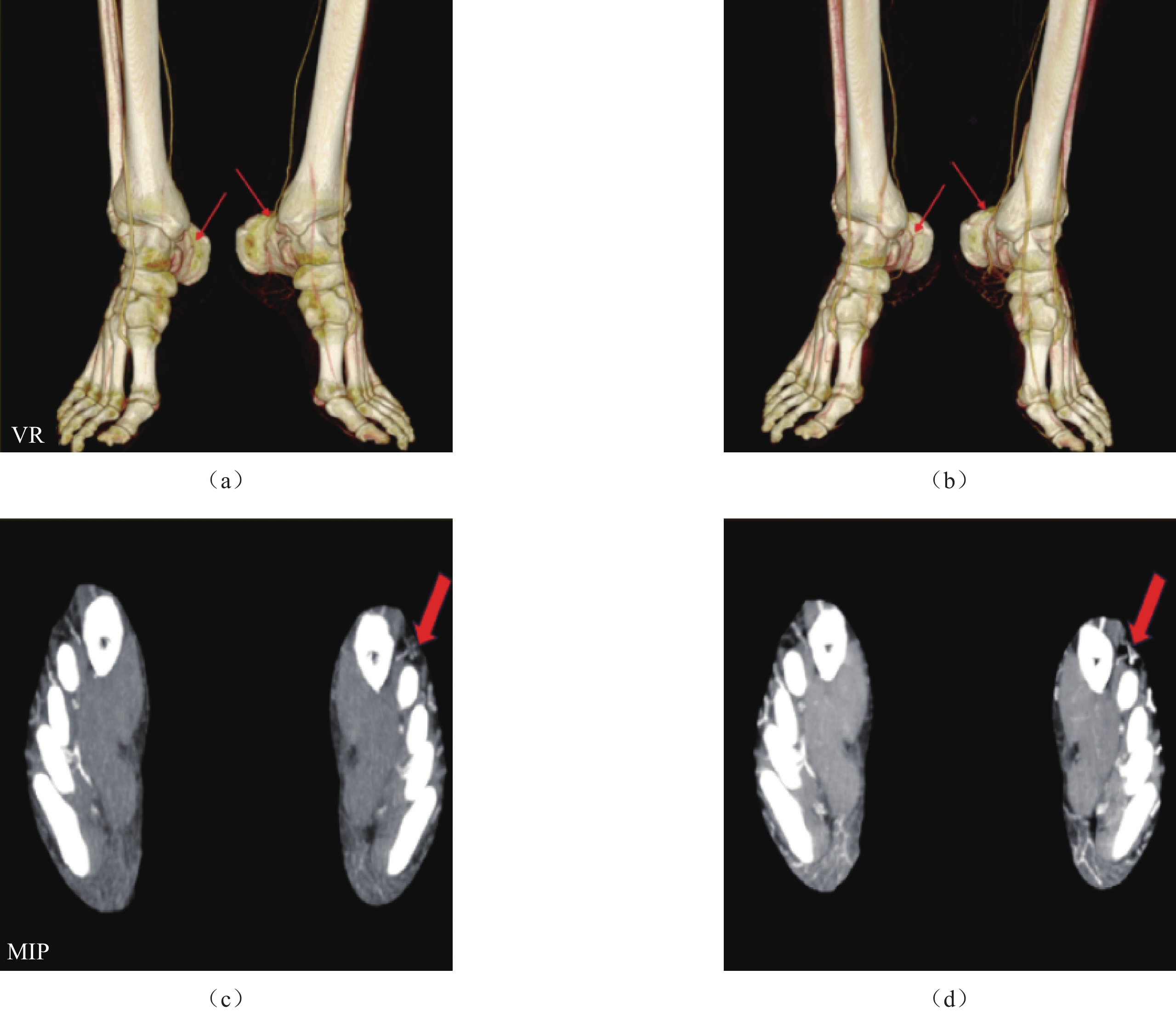
Objective: To investigate the feasibility of dual-energy computed tomography (DECT) in foot arteriography for small vessel image quality in patients with diabetes . Methods: A total of 30 patients with diabetes were enrolled prospectively between January and November 2022 in the radiology department of our hospital, where DECT was acquired immediately after the CT angiography (routine CTA group) of the lower extremity.The optimal virtual monochromatic images (DECT group) were derived from the DECT data. Image quality was assessed by measuring the CT values and noise in foot arteries, and signal-tonoise ratio (SNR) and contrast-to-noise ratio (CNR) were calculated. The arterial course and small vessel display were scored using a 5-point scale (1, poor; 5, excellent). Results: The CNR and SNR were significantly better in the DECT group than in the routine CTA group. The DECT group received higher subjective scores on the posterior tibial artery and the foot arteries (all > 3) than did the routine CTA group. Conclusion: Compared to the routine CTA imaging, DECT offers superior image quality for foot arteriography, thereby enhancing the diagnostic accuracy of foot arteriography and bolstering physicians’ confidence in disease diagnosis.
| [1] |
MISHRA S C, CHHATBAR K C, KASHIKAR A, et al. Diabetic foot[J]. British Medical Journal, 2017, 359(S1): 1-7.
|
| [2] |
纪东华, 刘暴. 膝下动脉血运重建的临床实践中国专家共识[J]. 中国循环杂志, 2024, 39(2): 116-122. DOI: 10.3969/j.issn.1000-3614.2024.02.002.
JI D H, LIU B. Chinese expert consensus on the standards of practice on below-the-knee arteries revascularisation[J]. Chinese Circulation Journal, 2024, 39(2): 116-122. DOI: 10.3969/j.issn.1000-3614.2024.02.002. (in Chinese).
|
| [3] |
孙旭, 王东军, 朱朝军, 等. 中医外治视域下糖尿病足研究关键问题及思考[J]. 时珍国医国药, 2023, 34(12): 2984-2986.
|
| [4] |
COLE J B, FLOREZ J C. Genetics of diabetes mellitus and diabetes complications[J]. Nature Review Nephrology, 2020, 16(7): 377-390. DOI: 10.1038/s41581-020-0278-5.
|
| [5] |
FORBES J M, COOPER M E. Mechanisms of diabetic complications[J]. Physiological Review, 2013, 93(1): 137-188. DOI: 10.1152/physrev.00045.2011.
|
| [6] |
CORRIERE M, ROOPARINESINGH N, KALYANI R R. Epidemiology of diabetes and diabetes complications in the elderly: An emerging public health burden[J]. Current Diabetes Report, 2013, 13(6): 805-813. DOI: 10.1007/s11892-013-0425-5.
|
| [7] |
SHARMA S, SCHAPER N, RAYMAN G. Microangiopathy: Is it relevant to wound healing in diabetic foot disease?[J]. Diabetes/Metabolism Reserch and Review, 2020, 36(S1): e3244. DOI: 10.1002/dmrr.3244.
|
| [8] |
郭晓曦, 林禹, 吕绍茂, 等. 下肢动脉双能量CT成像不同能量融合图像与单能量图像的对比研究[J]. 临床放射学杂志, 2021, 40(2): 363-368.
GUO X X, LIN Y, LV S M, et al. A comparative study of different fusion image and monoenergetic image in dual-energy CT of lower extremity artery[J]. Journal of Clinical Radiology, 2021, 40(2): 363-368. (in Chinese).
|
| [9] |
刘衡, 冉启胜, 夏传江, 等. 下肢动脉血管造影技术对糖尿病足患者末梢动脉准确显示的应用研究[J]. 重庆医科大学学报, 2019, 44(10): 1351-1354.
LIU H, RAN Q S, XIA C J, et al. Application of CT-angiography in accurate display of peripheral arteries in patients with diabetic foot[J]. Journal of Chongqing Medical University, 2019, 44(10): 1351-1354. (in Chinese).
|
| [10] |
HSIEH J, FLOHR T. Computed tomography recent history and future perspectives[J]. Journal of Medical Imaging, 2021, 8(5): 52109.
|
| [11] |
MCCOLLOUGH C H, LENG S, YU L, et al. Dual-and multi-energy CT: Principles, technical approaches, and clinical applications[J]. Radiology, 2015, 276(3): 637-653. DOI: 10.1148/radiol.2015142631.
|
| [12] |
JACOBSEN M C, THROWER S L, GER R B, et al. Multi-energy computed tomography and material quantification: Current barriers and opportunities for advancement[J]. Journal of Medical Physics, 2020, 47(8): 3752-3771. DOI: 10.1002/mp.14241.
|
| [13] |
王文杰, 陈平, 潘晋孝, 等. 基于参考组分的双能CT成像方法[J]. CT理论与应用研究, 2021, 30(1): 61-69. DOI: 10.15953/j.1004-4140.2021.30.01.06.
WANG W J, CHEN P, PAN J X, et al. Dual-energy CT imaging method based on reference components[J]. CT Theory and Applications, 2021, 30(1): 61-69. DOI:10.15953/j.1004-4140.2021.30.01.06. (in Chinese).
|
| [14] |
梁洪伟, 周旸, 张志伟, 等. 双能CT虚拟单能级成像显示胰周血管: 与线性融合图像对照研究[J]. 中国医学计算机成像杂志, 2022, 28(1): 56-62. DOI: 10.3969/j.issn.1006-5741.2022.01.012.
LIANG H W, ZHOU Y, ZHANG Z W, et al. Dual-energy CT with virtual monoenergetic images in demonstration of peripancreatic vessels: Comparison with polyenergetic images[J]. Chinese Computed Medical Imaging, 2022, 28(1): 56-62. DOI: 10.3969/j.issn.1006-5741.2022.01.012. (in Chinese).
|
| [15] |
侯凯, 吕鹏, 顾君英, 等. 实时阈值手动触发技术在下肢动脉CT血管成像中的临床应用价值[J]. 中国临床医学, 2016, 23(1): 81-85.
HOU K, LV P, GU J Y, et al. The clinical application value of real time threshold manual trigering technique in the lower extremity arterial CT angiography[J]. Chinese Journal of Clinical Medicine, 2016, 23(1): 81-85. (in Chinese).
|
| [16] |
WICHMANN J L, GILLOTT M R, de CECCO C N, et al. Dual-energy computed tomography angiography of the lower extremity runoff: Impact of noiseoptimized virtual monochromatic imaging on image quality and diagnostic accuracy[J]. Investigative Radiology, 2016, 51(2): 139-146. DOI: 10.1097/RLI.0000000000000216.
|
| [17] |
JIA X, LI X, LI J, et al. Improving diagnostic accuracy for arteries of lower extremities with dual-energy spectral CT imaging[J]. European Journal of Radiology, 2020, 128: 109061. DOI: 10.1016/j.ejrad.2020.109061.
|
| [18] |
WANG G, ZHAO D, LING Z, et al. Evaluation of the best single-energy scanning in energy spectrum CT in lower extremity arteriography[J]. Experimental and Therapeutic Medicine, 2019, 18(2): 1433-1439.
|
| [19] |
SUN H, SAEEDI P, KARURANGA S, et al. IDF diabetes atlas: Global, regional and country-level diabetes prevalence estimates for 2021 and projections for 2045[J/OL]. Diabetes Research and Clinical Practice, 2022, 183: 109-119.
|
| [20] |
JUDE E B, OYIBO S O, CHALMERS N, et al. Peripheral arterial disease in diabetic and nondiabetic patients: A comparison of severity and outcome[J]. Diabetes Care, 2001, 24(8): 1433-1437. DOI: 10.2337/diacare.24.8.1433.
|
| [21] |
GOLLEDGE J. Update on the pathophysiology and medical treatment of peripheral artery disease[J]. Nature Reviews Cardiology, 2022, 19(7): 456-474. DOI: 10.1038/s41569-021-00663-9.
|
| [22] |
LOW WANG C C, BLOMSTER J I, HEIZER G, et al. Cardiovascular and limb outcomes in patients with diabetes and peripheral artery disease: The EUCLID Trial[J]. Journal of the American College of Cardiology, 2018, 72(25): 3274-3284. DOI: 10.1016/j.jacc.2018.09.078.
|
| [23] |
任宁, 李启富, 程庆丰. 糖尿病周围动脉疾病的诊疗进展[J]. 重庆医学, 2012, 41(33): 3547-3549. DOI: 10.3969/j.issn.1671-8348.2012.33.037.
|
| [24] |
American Diabetes Association. Microvascular complications and foot care: Standards of medical care in diabetes[J]. Diabetes Care, 2018, 41(S1): S105-S118.
|
| [1] | LI Guorong, SUN Yang, ZHANG Tingting. Advances in the Use of CMR in Subclinical Diabetic Cardiomyopathy[J]. CT Theory and Applications, 2023, 32(6): 836-842. DOI: 10.15953/j.ctta.2022.255 |
| [2] | LIANG Yuhong, ZHONG Xixi, YAO Xinqun, HUANG Shiqi, LUO Lian, LYU Yaping. High-resolution Computed Tomography (HRCT) Characteristics of Coronavirus Disease 2019 (COVID-19) in Patients with Diabetes[J]. CT Theory and Applications, 2023, 32(5): 659-665. DOI: 10.15953/j.ctta.2023.031 |
| [3] | LI Ling, ZHANG Mingxia, SUN Ying, DUAN Shuhong, GUO Jia, DU Changyue, LIU Mengke, ZHANG Yimeng, SUN Lei, HUO Meng, WANG Rengui. Imaging Study of COVID-19 Patients with Diabetes Mellitus by Computed Tomograpgh Quantitative Indicators Based on Deep Learning[J]. CT Theory and Applications, 2023, 32(3): 373-379. DOI: 10.15953/j.ctta.2023.020 |
| [4] | ZHANG Fengling, ZHAO Li, LIU Jiabao. CT Features of Pulmonary Infection in Elderly Patients with Type 2 Diabetes Mellitus[J]. CT Theory and Applications, 2021, 30(5): 583-590. DOI: 10.15953/j.1004-4140.2021.30.05.06 |
| [5] | LAI Tianfu, DENG Junliang, CHEN Xiangguang, YAO Chun. Analysis of CTA Characterisation of Coronary Atherosclerosis with Diabetes Mellitus[J]. CT Theory and Applications, 2020, 29(3): 347-354. DOI: 10.15953/j.1004-4140.2020.29.03.11 |
| [6] | SUN Pengtao, DOU Xuechao, SUN Xiaoli, ZHAO Tong, WEI Hailiang, WEN Tingguo, WANG Rengui. The Relationship between Pancreatic Steatosis and Framingham Cardiovascular Risk Score in Patients with Type 2 Diabetes Mellitus[J]. CT Theory and Applications, 2019, 28(4): 463-470. DOI: 10.15953/j.1004-4140.2019.28.04.07 |
| [7] | HE Wei-hong, FU Xi, KE Qi, YUAN Jian-xiang, DONG Xiang-yu. Analysis of 64 Slice Spiral CT Angiography in Diabetic Lower Extremity Arterial Disease[J]. CT Theory and Applications, 2018, 27(3): 373-378. DOI: 10.15953/j.1004-4140.2018.27.03.10 |
| [8] | XIE Wan-meng, CHEN Jun, WANG Jun, XIE Xin-jia. A Primary Study of Magnetic Resonance Imaging-diffusion Weighted Imaging of Pancreas in Type 2 Diabetic Mellitus Patients[J]. CT Theory and Applications, 2016, 25(5): 563-569. DOI: 10.15953/j.1004-4140.2016.25.05.08 |
| [9] | YANG Chun-yu, SHEN Bi-xian, ZHAO Yue, HUANG Yin-ping, CHEN Sheng-ji, HUANG An-rong. Study on the Value of Dual Source CT Assessment of Correlation between Diabetes and Coronary Plaque[J]. CT Theory and Applications, 2014, 23(6): 913-921. |
| [10] | ZHANG Yong-dong. Phthisical CT Diagnosis in Diabetes(with 22 Cases Report)[J]. CT Theory and Applications, 2006, 15(1): 33-36. |
Supported by: Beijing Renhe Information Technology Co. Ltd 


 DownLoad: