ISSN 1004-4140
CN 11-3017/P

| Citation: |
WEI P Y, HUANG X Y, WU T, et al. Effect of denture materials on CT image artifacts: phantom experiment[J]. CT Theory and Applications, xxxx, x(x): 1-7. DOI: 10.15953/j.ctta.2024.339. (in Chinese).

|
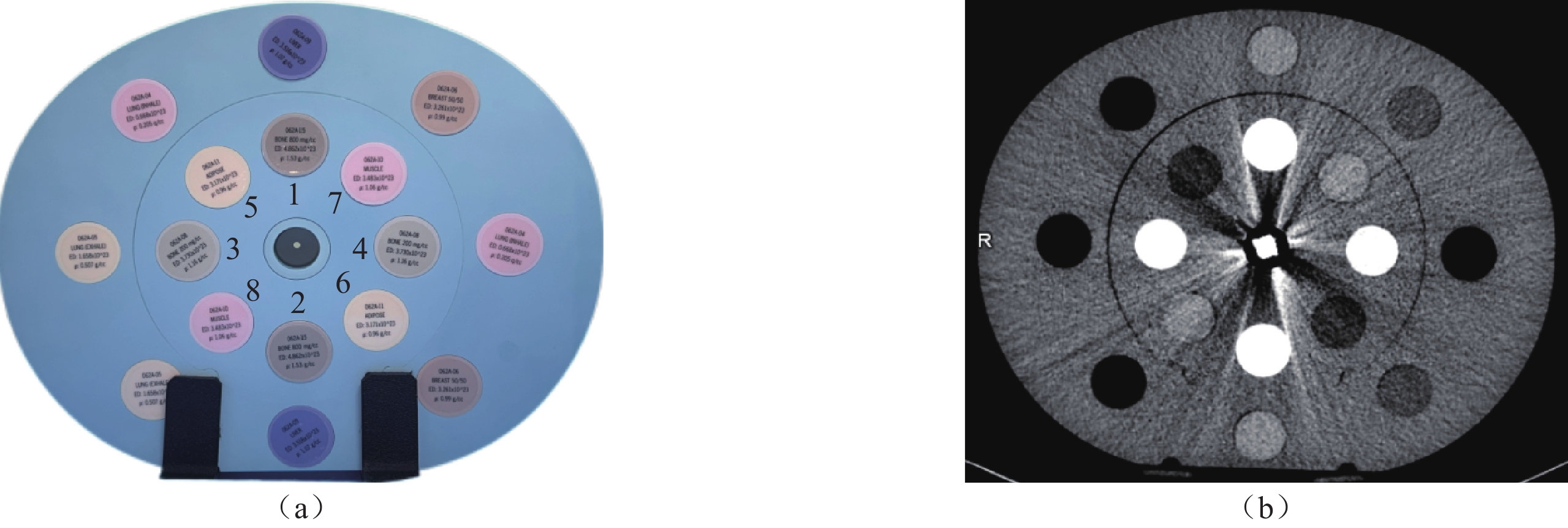
Objective: Based on phantom experiments, this study aims to optimize artifacts in multimaterial dentures and provide guidance for clinical practice. Method: A blank control and six denture full-crown models of different materials are placed sequentially at the CIRS phantom center. Subsequently, dual-energy CT scanning is performed using energy CT, and the optimal contrast image of the blank control is reconstructed as a baseline. Next, the optimal contrast images are reconstructed under standard and bone algorithms for the six groups, in addition to virtual monochromatic images at 70–140 keV with an interval of 10 keV. CT and SD values are recorded, and the artifact index (AI) is calculated. Routine scanning is performed and radiation doses of the routine and energy groups are recorded. Two radiologists subjectively evaluate the images of each group and perform a variance analysis or nonparametric tests for data analysis. Results: The radiation dose of the energy group (6.15±0.10) mGy is significantly lower than that of the routine group (30.35±0.02) mGy. The consistency of the subjective image scoring is favorable. As the energy levels increase, the image quality of the energy group improves, whereas the AI decreases. Standard algorithm: The AI values of all modules except the upper fat differ significantly. The AI values of all modules except the light bone differ significantly because of the different denture materials used. Bone algorithm: The AI values of all modules differ significantly, and the AI values of fat and muscle differ significantly owing to the different denture materials. Conclusion: Energy CT significantly reduces the radiation dose. Additionally, pure-titanium dentures have the least number of artifacts and are the preferred material for clinical full crowns. Energy CT combined with virtual single-energy imaging can reduce artifacts and optimize image quality. Recommendations for clinical observation of bone tissue are as follows: Pure titanium, 140 keV; cobalt–chromium, 90 keV; cobalt–chromium porcelain, 140 keV; nickel–chromium, 120keV; nickel–chromium porcelain, 120 keV; and all ceramic, 140 keV.
| [1] |
崔永斌, 黄勇, 李若影, 等. 光谱CT对头颈部金属植入物伪影的去除作用: 一项基于人仿真模体的研究[J]. 临床放射学杂志, 2023, 42(12): 1997-2006. DOI: 10.13437/j.cnki.jcr.2023.12.019.
CUI Y B, HUANG Y, LI R Y, et al. Reduction of artifacts from head and neck metal implants by spectral CT: A study based on human simulation phantom[J]. Journal of Clinical Radiology, 2023, 42(12): 1997-2006. DOI: 10.13437/j.cnki.jcr.2023.12.019.
|
| [2] |
孙强, 牛志兴, 郑宏雨, 等. 多物质伪影降低技术对五种口腔修复材料能谱CT伪影消除作用初探[J]. 中华口腔医学杂志, 2019, 54(11): 760-764. DOI: 10.3760/cma.j.issn.1002?0098.2019.11.007.
SUN Q, NIU Z X, ZHENG H Y, et al. Preliminary study on the elimination of artifacts of five kinds of dental prosthetic materials by energy spectrum CT multi-material artifact reduction technology[J]. Chinese Journal of Stomatology, 2019, 54(11): 760-764. DOI: 10.3760/cma.j.issn.1002?0098.2019.11.007.
|
| [3] |
KITAMI R, IZUMI M, TANIGUCHI M, et al. Phantom study for CT artifacts of dental titanium implants and zirconia upper structures: the effects of occlusal plane angle setting and SEMAR algorithm[J]. Oral Radiology, 2024, Apr;40(2): 251-258. DOI: 10.1007/s11282-023-00730-6.
|
| [4] |
FOTI G, ASCENTI G, AGOSTINI A, et al. Dual-energy CT in oncologic imaging[J]. Tomography (Ann Arbor, Mich. ), 2024, 23;10(3): 299-319. DOI: 10.3390/tomography10030024.
|
| [5] |
周洁. 第3代双源CT虚拟单能量成像减少口腔金属伪影的效果评估[D]. 太原: 山西医科大学, 2021. DOI: 10.27288/d.cnki.gsxyu.2021.000498.
ZHOU J. To Evaluate the effect of the third-generation dual-source CT virtual monoenergetic reconstruction technique in reducing oral metal artifacts[D]. Tai Yuan: Shanxi Medical University, 2021. DOI:10.27288/d.cnki.gsxyu.2021.000498.(in Chinese).
|
| [6] |
中华医学会放射学分会, 中国医师协会放射医师分会, 安徽省影像临床医学研究中心. 能量CT临床应用中国专家共识[J]. 中华放射学杂志, 2022, 56(05): 476-487. DOI: 10.3760/cma.j.cn112149-20220118-00051.
|
| [7] |
HENZLER T, FINK C, SCHOENBERG S O, et al. Dual-energy CT: radiation dose aspects[J]. American journal of roentgenology, 2012, 199(5 Suppl): S16-25. DOI: 10.2214/AJR.12.9210.
|
| [8] |
PALLASCH F B, RAU A, REISERT M, et al. Impact of different metal artifact reduction techniques in photon-counting computed tomography head and neck scans in patients with dental hardware[J]. European Radiology, 2024, 34(6): 3742-3749. DOI: 10.1007/s00330-023-10430-8.
|
| [9] |
高志远, 隋岩, 刘康, 等. 基于特定条件下头颅模体螺旋扫描与轴位扫描的图像质量和辐射剂量的对比研究[J]. 中国医学装备, 2024, 21(09): 18-22. DOI: 10.3969/j.issn.1672-8270.2024.09.004.
GAO Z Y, SUI Y, LIU K, et al. Comparative study on image quality and radiation dose between the spiral scanning and the axial scanning for skull phantom based onspecific conditions[J]. China Medical Equipment, 2024, 21(09): 18-22. DOI: 10.3969/j.issn.1672-8270.2024.09.004.
|
| [10] |
蒋东, 秦立新. 能谱纯化Sn 150 kV结合ADMIRE在腰椎CT检查中的价值[J]. CT理论与应用研究(中英文), 2024, 33(1): 49-55. DOI: 10.15953/j.ctta.2022.240.
JIANG D, QIN L X. Spectral Filtration Sn 150 kV Combined with Advanced Simulated Iterative Reconstruction in Lumbar Computed Tomography Examination[J]. CT Theory and Applications, 2024, 33(1): 49-55. DOI: 10.15953/j.ctta.2022.240.
|
| [11] |
孙琦, 董敏俊, 杨星, 等. 口腔颌面部CT能谱成像降低金属伪影的效果评价[J]. 上海口腔医学, 2017, 26(06): 646-649. doi: 10.19439/j.sjos.2017.06.016
SUN Q, DONG M J, YANG X, et al. Clinical analysis of spectrum CT imaging reducing metal artifacts of oral and maxillofacial region.[J]. Shanghai Journal of Stomatology, 2017, 26(06): 646-649. doi: 10.19439/j.sjos.2017.06.016
|
| [12] |
甘露, 刘基, 袁晨, 等. 能谱CT结合MARs技术对不同材质义齿伪影去除的临床价值[J]. 天津医药, 2023, 51(6): 642-647. DOI: 10.11958/20221330.
GAN L, LIU J, YUAN C, et al. Clinical value of energy spectrum CT combined with MARs technique for artifact removal of dentures of different materials[J]. Tianjin Medical Journal, 2023, 51(6): 642-647. DOI: 10.11958/20221330.
|
| [13] |
李霞敏, 黄晓颖, 李知非, 等. 双源Mono+技术联合实影渲染技术在提高肝硬化门静脉成像中的应用[J]. CT理论与应用研究(中英文), 2025, 34(1): 44-50. DOI: 10.15953/j.ctta.2024.149.
LI X M, HUANG X Y, LI Z F, et al. Application of Dual Source Mono+Combined with Cinematic Rendering in Cirrhosis Portal Vein Imaging[J]. CT Theory and Applications, 2025, 34(1): 44-50. DOI: 10.15953/j.ctta.2024.149.
|
| [14] |
马月, 何长久, 胡仕北, 等. 双能量CT虚拟平扫在颈部扫描中的应用价值[J]. 肿瘤预防与治疗, 2024, 37(2): 156-161. DOI: 10.3969/j.issn.1674-0904.2024.02.008.
MA Y, HE C J, HU S B, et al. Application Value of Virtual Non-contrast Dual-Energy CT in Neck CT Examination[J]. Journal of Cancer Control and Treatment, 2024, 37(2): 156-161. DOI: 10.3969/j.issn.1674-0904.2024.02.008.
|
| [15] |
杨亮, 罗德红, 赵燕风, 等. 头颈部肿瘤检查中能谱CT虚拟平扫替代常规平扫的可行性研究[J]. 中华放射学杂志, 2015(8): 572-576. DOI: 10.3760/cma.j.issn.1005-1201.2015.08.003.
YANG L, LUO D H, ZHAO Y F, et al. Feasibility study on application of gemstone spectral CT material suppressed iodine as virtual non-contrast CT scan in head and neck neoplasms[J]. Chinese Journal of Radiology, 2015(8): 572-576. DOI: 10.3760/cma.j.issn.1005-1201.2015.08.003.
|
| [16] |
李文超, 李强, 李晨光, 等. 能谱CT最佳单能量技术、多模型迭代重建算法及虚拟平扫联合应用在头颈CTA应用研究[J]. 中国CT和MRI杂志, 2023, 21(12): 38-41. DOI: 10.3969/j.issn.1672-5131.2023.12.012.
LI W C, LI Q, LI C G, et al. Application Study of the Combined Application of Spectral CT Optimal Single Energy Technique, Adaptive Statistical Iterative Reconstruction-V and Virtual Plain Scan in Head and Neck CTA[J]. Chinese Journal of CT and MRI, 2023, 21(12): 38-41. DOI: 10.3969/j.issn.1672-5131.2023.12.012.
|
| [17] |
黄琼, 吴梦雄, 董海鹏, 等. 基于骨重建算法结合ASIR-V在冠状动脉支架成像中的应用研究[J]. 诊断学理论与实践, 2022, 21(01): 68-73. DOI: 10.16150/j.1671-2870.2022.01.013.
HUANG Q, WU M X, DONG H P, et al. Study on application of bone algorithm combined with ASIR-V in coronary stent imaging[J]. Journal of Diagnostics Concepts & Practice, 2022, 21(01): 68-73. DOI: 10.16150/j.1671-2870.2022.01.013.
|
| 1. |
张科,张春晓. 基于深度残差网络的儿科肺炎辅助诊断算法. 中国医疗设备. 2022(09): 42-46+56 .

| |
| 2. |
周丽媛,赵启军,高定国. 基于注意力引导深度纹理特征学习的复杂背景藏药材切片图像识别. 世界科学技术-中医药现代化. 2022(12): 4825-4832 .
|
Supported by: Beijing Renhe Information Technology Co. Ltd 


 DownLoad: