ISSN 1004-4140
CN 11-3017/P
| Citation: |
YAN X, ZHAO J H. Computed Tomography and Magnetic Resonance Imaging Diagnosis of Intracranial Solitary Fibrous Tumor: A Clinical Case Analysis[J]. CT Theory and Applications, 2024, 33(3): 365-370. DOI: 10.15953/j.ctta.2023.126. (in Chinese).

|
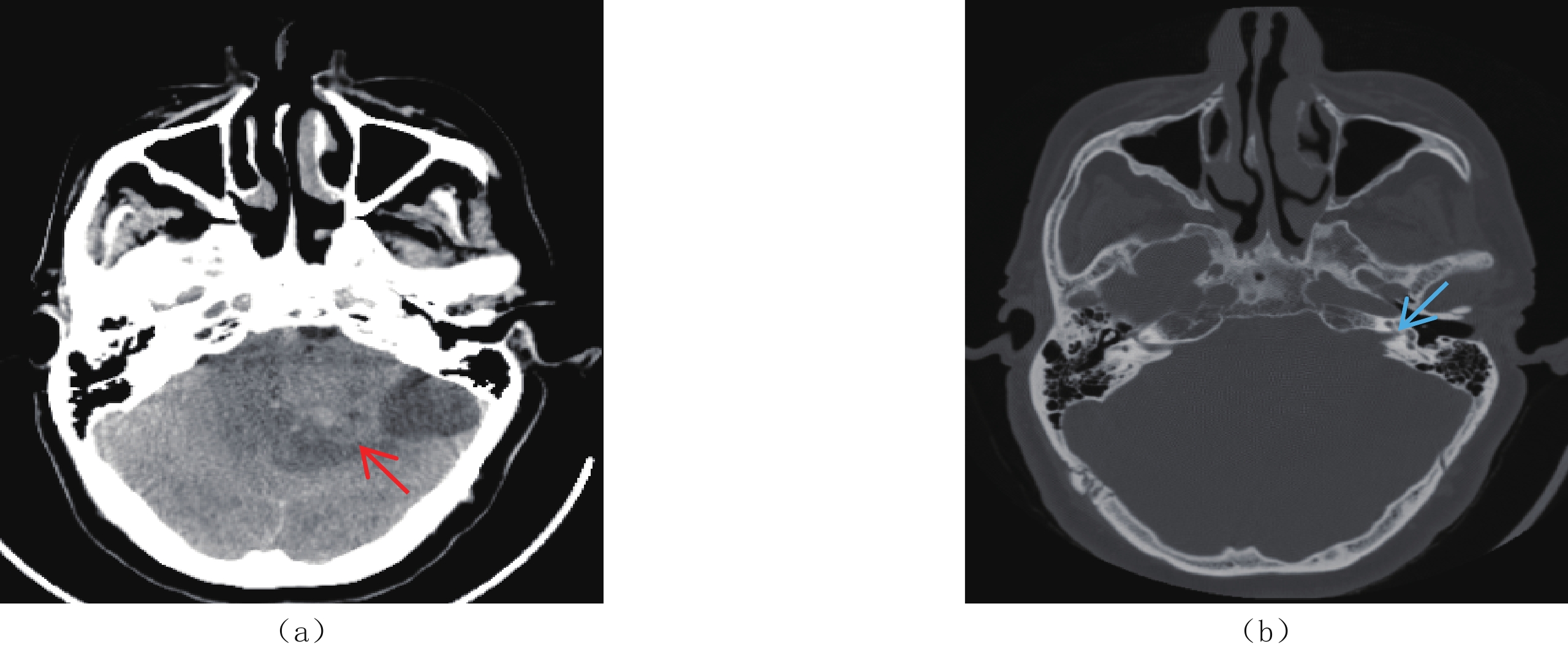
Intracranial solitary fibroma tumor (ISFT) is a kind of mesenchymal tissue-derived spindle cell tumor. Patients’ disease progression vary according to the course and location of the tumor. Due to its rarity, radiologists and clinicians lack a comprehensive understanding of ISFT. Hence, the preoperative misdiagnosis rate is high. This case report describes a 52-year-old male patient with intracranial solitary fibroma who presented with headache and weakness of both lower extremities. He underwent radiological examination, including computed tomography (CT) and magnetic resonance imaging (MRI), and was diagnosed with acoustic neuroma. After postoperative pathological tissue biopsy, he was diagnosed with ISFT. He was reviewed one and a half years after surgery; there was no significant discomfort in addition to the paralysis of the left facial nerve and swelling of the left side of the face. This case report retrospectively analyzes the radiological scans and the clinical data of the patient to summarize the key CT and MRI features of ISFT, improving the accuracy of the preoperative diagnosis of this rare disease, and contributing to current knowledge of the precise treatment of ISFT.
| [1] |
CARNEIRO S S, SCHEITHAUER B W, NASCIMENTO A G, et al. Solitary fibrous tumor of the meninges: A lesion distinct from fibrous meningioma. A clinicopathologic and immunohistochemical study[J]. American Journal of Clinical Pathology, 1996, 106(2): 217−224. DOI: 10.1093/ajcp/106.2.217.
|
| [2] |
LOUIS D N, PERRY A, WESSELING P, et al. The 2021 WHO classification of tumors of the central nervous system: A summary[J]. Neuro-Oncology, 2021, 23(8): 1231−1251. DOI: 10.1093/neuonc/noab106.
|
| [3] |
吴虹林, 苏伟杰, 李西西, 等. 中枢神经系统孤立性纤维瘤临床特征及术后复发因素[J]. 中国神经精神疾病杂志, 2023,49(2): 85−91. DOI: 10.3969/j.issn.1002-0152.2023.02.003.
WU H J, SU W J, LI X X, et al. Clinical features and recurrent factors of solitary fibrous tumor in central nervous system[J]. Chinese Journal of Nervous and Mental Diseases, 2023, 49(2): 85−91. DOI: 10.3969/j.issn.1002-0152.2023.02.003. (in Chinese).
|
| [4] |
MIETTINEN M M, EL-RIFAI W, SARLOMO-RIKALA M, et al. Tumor size-related DNA copy number changes occur in solitary fibrous tumors but not in hemangiopericytomas[J]. Modern Pathology, 1997, 10(12): 1194−1200.
|
| [5] |
FARGEN K M, OPALACH K J, WAKEFIELD D, et al. The central nervous system solitary fibrous tumor: A review of clinical, imaging and pathologic findings among all reported cases from 1996 to 2010[J]. Clinical Neurology and Neurosurgy, 2011, 113(9): 703−710. DOI: 10.1016/j.clineuro.2011.07.024.
|
| [6] |
高文华, 黄玉芳, 朱小贵, 等. 颅内孤立性纤维瘤影像表现[J]. 现代医用影像学, 2019,28(7): 1472−1474. doi: 10.3969/j.issn.1006-7035.2019.07.005
GAO W H, HUANG Y F, ZHU X G, et al. Imaging characters of intracranial solitary fibrous tumor[J]. Modern Medical Imageology, 2019, 28(7): 1472−1474. (in Chinese). doi: 10.3969/j.issn.1006-7035.2019.07.005
|
| [7] |
梅磊磊, 聂蕾, 唐文英, 等. 孤立性纤维瘤的影像表现及临床病理特征[J]. 放射学实践, 2022,37(5): 566−570. DOI: 10.13609/j.cnki.1000-0313.2022.05.006.
MEI L L, NIE L, TANG W Y, et al. Imaging and clinicopathologic features of solitary fibrous tumor[J]. Radiologic Practice, 2022, 37(5): 566−570. DOI: 10.13609/j.cnki.1000-0313.2022.05.006. (in Chinese).
|
| [8] |
曾禹莉, 易熙, 赵洁, 等. 颅内孤立性纤维瘤的CT及MRI表现[J]. 影像研究与医学应用, 2022,6(4): 163−165. doi: 10.3969/j.issn.2096-3807.2022.04.055
|
| [9] |
CLARENCON F, BONNEVILLE F, ROUSSEAU A, et al. Intracranial solitary fibrous tumor: Imaging findings[J]. European Journal of Radiology, 2011, 80(2): 387−394. DOI: 10.1016/j.ejrad.2010.02.016.
|
| [10] |
何文杰, 雷益, 焦娟, 等. 颅内孤立性纤维瘤/血管外皮瘤的影像表现与病理分析[J]. 放射学实践, 2019,34(12): 1299−1303. DOI: 10.13609/j.cnki.1000-0313.2019.12.004.
HE W J, LEI Y, JIAO J, et al. Imaging features and pathological analysis of intracranial solitary fibrous tumor/hemangiopericytoma[J]. Radiologic Practice, 2019, 34(12): 1299−1303. DOI: 10.13609/j.cnki.1000-0313.2019.12.004. (in Chinese).
|
| [11] |
冯加和, 杨云竣, 杨粤龙, 等. 颅内孤立性纤维瘤/血管外皮瘤瘤内血管流空特征研究[J]. 医学影像学杂志, 2022,32(8): 1273−1276. doi: 10.3969/j.issn.1006-9011.2022.8.yxyxxzz202208002
FENG J H, YANG Y J, YANG Y L, et al. Study on the characteristics of intratumoral vascular flow void in solitary fibrous tumor/hemangiopericytoma[J]. Journal of Medical Imaging, 2022, 32(8): 1273−1276. (in Chinese). doi: 10.3969/j.issn.1006-9011.2022.8.yxyxxzz202208002
|
| [12] |
张宇泽, 杨云竣, 陈静勿, 等. 血管瘤型脑膜瘤与颅内孤立性纤维瘤的MRI征象分析[J]. 临床放射学杂志, 2022,41(11): 2010−2014. DOI: 10.13437/j.cnki.jcr.2022.11.007.
ZHANG Y Z, YANG Y J, CHEN J W, et al. A comparative study on MRI signs between angiomatous meningioma and solitary fibrous tumor[J]. Journal of Clinical Radiology, 2022, 41(11): 2010−2014. DOI: 10.13437/j.cnki.jcr.2022.11.007. (in Chinese).
|
| [13] |
陶静雄, 梁奕, 王佳, 等. 颅内孤立性纤维瘤的影像学和病理学特征[J]. 医学影像学杂志, 2019,29(11): 1976−1978.
TAO J X, LIANG Y, WANG J, et al. Imaging and pathological features of intracranial solitary fibrous tumor[J]. Journal of Medical Imaging, 2019, 29(11): 1976−1978. (in Chinese).
|
| [14] |
柯代波, 刘文科, 张思, 等. 颅内孤立性纤维瘤的MRI表现[J]. 华西医学, 2017,32(1): 46−50. DOI: 10.7507/1002-0179.201511166.
KE D B, LIU W K, ZHANG S, et al. MRI manifestations of intracranial solitary fibrous tumor[J]. West China Medical Journal, 2017, 32(1): 46−50. DOI: 10.7507/1002-0179.201511166. (in Chinese).
|
| [15] |
DOYLE L A, VIVERO M, FLETCHER C D, et al. Nuclear expression of STAT6 distinguishes solitary fibrous tumor from histologic mimics[J]. Modern Pathology, 2014, 27(3): 390−395. DOI: 10.1038/modpathol.2013.164.
|
| [1] | ZHANG Ran, KONG Huihua, LI Jiaxin, SONG Yijiao. Dense Sandstone Material Decomposition Based on Improved Convolutional Neural Network[J]. CT Theory and Applications, 2025, 34(1): 117-128. DOI: 10.15953/j.ctta.2024.131 |
| [2] | Du Congcong, Qiao Zhiwei. EPRI Sparse-Reconstruction Method Based on Deep Learning[J]. CT Theory and Applications. DOI: 10.15953/j.ctta.2025.047 |
| [3] | QIAO Yiyu, QIAO Zhiwei. Low-dose CT Image Reconstruction Method Based on CNN and Transformer Coupling Network[J]. CT Theory and Applications, 2022, 31(6): 697-707. DOI: 10.15953/j.ctta.2022.114 |
| [4] | HAN Zefang, SHANGGUAN Hong, ZHANG Xiong, HAN Xinglong, GUI Zhiguo, CUI Xueying, ZHANG Pengcheng. Advances in Research on Low-dose CT Imaging Algorithm Based on Deep Learning[J]. CT Theory and Applications, 2022, 31(1): 117-134. DOI: 10.15953/j.1004-4140.2022.31.01.14 |
| [5] | HUANG Dongyun, XIA Jun, LIN Yuwen, CHEN Jiakuan, CHEN Haibin. The Application Value of Accurate Diagnosis of CT Image of Skull Base Fractures based on Convolutional Neural Network[J]. CT Theory and Applications, 2021, 30(6): 769-776. DOI: 10.15953/j.1004-4140.2021.30.06.13 |
| [6] | CHEN Kang, DI Guidong, ZHANG Jiajia, ZHOU You, WU Yao, ZHANG Guangzhi. Reservoir Prediction Based on Improved U-Net Convolutional Neural Network[J]. CT Theory and Applications, 2021, 30(4): 403-416. DOI: 10.15953/j.1004-4140.2021.30.04.01 |
| [7] | LIU Jiang, TU Qicui, LI Bingying, HUANG Xin, ZHANG Guangzhi, ZHANG Jiajia, WU Yao, ZHU Shengwei. Fault Prediction Method Based on Convolutional Neural Network[J]. CT Theory and Applications, 2020, 29(5): 522-533. DOI: 10.15953/j.1004-4140.2020.29.05.02 |
| [8] | CAI Ning, WANG Shijie, CHEN Lujie, ZHANG Yikun, CHEN Yang, LUO Shouhua, GU Ning. Low-dose Micro-CT Imaging Method Based on Progressive Network Processing[J]. CT Theory and Applications, 2020, 29(4): 435-446. DOI: 10.15953/j.1004-4140.2020.29.04.06 |
| [9] | CHEN Mai-Lin, SUN Ying-Shi. Clinical Value of Spectral CT Imaging with Rapid Voltage Switching for Pulmonary Nodules[J]. CT Theory and Applications, 2019, 28(6): 701-708. DOI: 10.15953/j.1004-4140.2019.28.06.08 |
| [10] | QIAO Zhi-wei. On Implementation and Stability Analysis of Two Types of Finite Hilbert Transforms Used in Backprojection Filtration Algorithms[J]. CT Theory and Applications, 2014, 23(4): 561-568. |
| 1. |
邓扬,陆金琦,余信江,胡超. 一种结合弹性波CT正演模拟与钻孔注水法的防渗墙检测方法研究. 计算机测量与控制. 2024(03): 125-130 .

| |
| 2. |
何继善,李帝铨,胡艳芳,凌帆,王金海. 城市强干扰环境地下空间探测技术与应用. 工程地球物理学报. 2022(05): 559-567 .
|
Supported by: Beijing Renhe Information Technology Co. Ltd 


 DownLoad: