ISSN 1004-4140
CN 11-3017/P
| Citation: |
QI W W, CHENG J, CHEN C H, et al. Value of Low Tube Voltage Combined with Deep Learning Image Reconstruction Algorithm to Reduce Radiation Dose in Combined Thoracoabdominal Enhanced CT[J]. CT Theory and Applications, 2025, 34(3): 359-368. DOI: 10.15953/j.ctta.2025.001. (in Chinese).

|
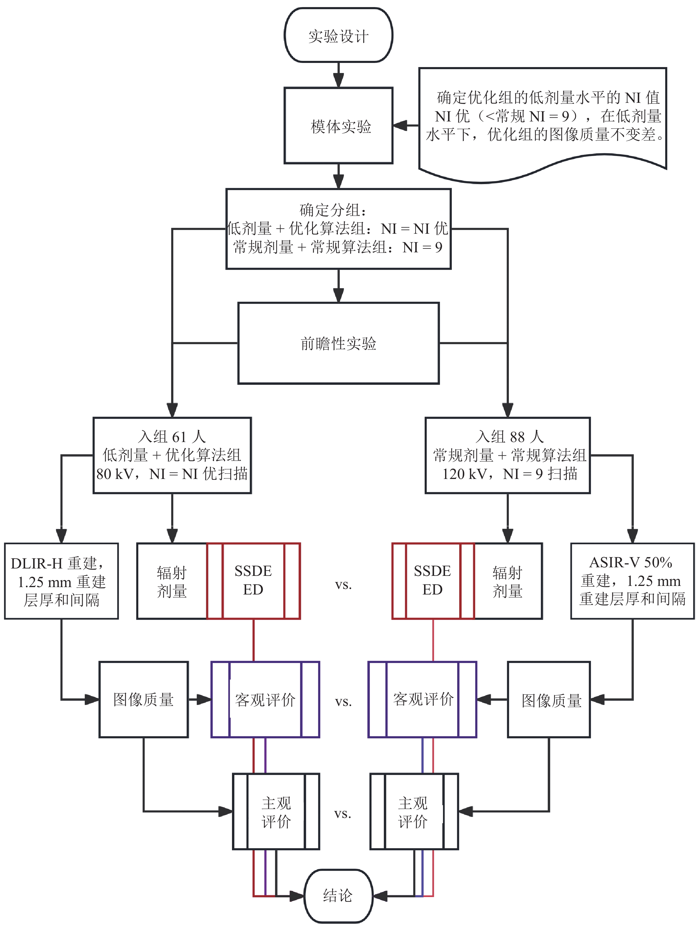
Objective: To investigate the effect of low tube voltage combined with deep learning image reconstruction (DLIR) on radiation dose reduction and maintaining image quality in combined chest and abdominal enhanced CT scans. Methods: (1) Phantom study. To determine the feasibility of combining low tube voltage with deep learning algorithms for low-contrast resolution, Catphan 500 phantoms were scanned under two different conditions. The optimization group used a low tube voltage (80 kV) combined with DLIR for scanning and image reconstruction, while the routine group used a 120 kV tube voltage combined with adaptive statistical iterative reconstruction V (ASiR-V). This study aimed to determine the effectiveness of the optimization group using a low dose (noise index, NI > 9) compared with the routine group using a routine dose (NI=9). (2) Prospective study. A total of 160 patients who underwent routine chest and abdominal enhanced CT scans were prospectively collected and randomly divided into a low-dose optimization group and routine-dose group, with 149 patients ultimately enrolled (61 in the low-dose optimization group and 88 in the routine-dose group). Based on the results of the phantom study, the low-dose optimization group used the optimized condition with NI set to the optimal value, whereas the routine-dose group used the routine condition with NI=9. Radiation doses were recorded and calculated for both groups, and image quality was subjectively and objectively evaluated. Results: The low-dose optimization group using NI=12 achieved an equivalent low-contrast resolution capability to the routine-dose group with NI=9. The effective dose in the low-dose optimization group (9.56±2.34) mSv was significantly lower than that in the routine-dose group (17.82±5.22) mSv. The liver and aorta attenuation values in the low-dose optimization group were significantly higher than those in the routine-dose group, and the CNR and SNR values in the liver and aorta were also significantly higher. The spatial resolution of the aorta, common hepatic artery, and portal vein and the display of small vessels/bronchi were all superior in the low-dose optimization group compared with the routine-dose group. Conclusion: The combination of a low tube voltage and deep learning image reconstruction algorithm can ensure equivalent or even higher image quality while reducing radiation dose, providing a feasible solution for optimizing radiation dose in large-scale CT scans such as the combined thoracoabdominal enhanced CT.
| [1] |
FURTADO C D, AGUIRRE D A, SIRLIN C B, et al. Whole-body CT screening: Spectrum of findings and recommendations in 1192 patients[J]. Radiology, 2005, 237(2): 385-394. DOI: 10.1148/radiol.2372041741.
|
| [2] |
BRANT-ZAWADZKI M. CT screening: Why I do it[J]. American Journal of Roentgenology, 2002, 179(2): 319-326. DOI: 10.2214/ajr.179.2.1790319.
|
| [3] |
SCHAUER D A, LINTON O W. National council on radiation protection and measurements report shows substantial medical exposure increase[J]. Radiology, 2009, 253(2): 293-296. DOI: 10.1148/radiol.2532090494.
|
| [4] |
REHANI M M, HEIL J, BALIYAN V. Multicentric study of patients receiving 50 or 100mSv in a single day through CT imaging-frequency determination and imaging protocols involved[J]. European Radiology, 2021, 31(9): 6612-6620. DOI: 10.1007/s00330-021-07734-y.
|
| [5] |
JENSEN C T, GUPTA S, SALEH M M, et al. Reduced-dose deep learning reconstruction for abdominal CT of liver metastases[J]. Radiology, 2022, 303(1): 90-98. DOI: 10.1148/radiol.211838.
|
| [6] |
刘方韬, 刘隺是, 陈勇, 等. 深度学习重建算法的图像质量体模研究[J]. CT理论与应用研究, 2022, 31(3): 351-356. DOI: 10.15953/j.ctta.2021.061.
LIU F T, LIU H S, CHEN Y, et al. Image quality assessment for deep learning image reconstruction algorithm: A phantom study[J]. CT Theory and Applications, 2022, 31(3): 351-356. DOI: 10.15953/j.ctta.2021.061. (in Chinese).
|
| [7] |
温德英, 杨杰尹, 汪琴, 等. 深度学习重建算法在上腹部CT成像中的应用[J]. CT理论与应用研究, 2022, 31(3): 329-336. DOI: 10.15953/j.ctta.2021.005.
WEN D Y, YANG J Y, WANG Q, et al. Application of deep learning reconstruction algorithm in upper abdomen CT[J]. CT Theory and Applications, 2022, 31(3): 329-336. DOI: 10.15953/j.ctta.2021.005. DOI: 10.15953/j.ctta.2021.005. (in Chinese).
|
| [8] |
NAM J G, HONG J H, KIM D S, et al. Deep learning reconstruction for contrast-enhanced CT of the upper abdomen: Similar image quality with lower radiation dose in direct comparison with iterative reconstruction[J]. European Radiology, 2021, 31(8): 5533-5543. DOI: 10.1007/s00330-021-07712-4.
|
| [9] |
LEE S, CHOI Y H, CHO Y J, et al. Noise reduction approach in pediatric abdominal CT combining deep learning and dual-energy technique[J]. European Radiology, 2021, 31(4): 2218-2226. DOI: 10.1007/s00330-020-07349-9.
|
| [10] |
KAWASHIMA H, ICHIKAWA K, TAKATA T, et al. Performance of clinically available deep learning image reconstruction in computed tomography: A phantom study[J]. Journal of Medical Imaging (Bellingham, Wash), 2020, 7(6): 63503.
|
| [11] |
LI L, WANG H, SONG J, et al. A feasibility study of realizing low-dose abdominal CT using deep learning image reconstruction algorithm[J]. Journal of X-ray Science and Technology, 2021, 29(2): 361-372.
|
| [12] |
CAO L, LIU X, LI J, et al. A study of using a deep learning image reconstruction to improve the image quality of extremely low-dose contrast-enhanced abdominal CT for patients with hepatic lesions[J]. British Journal of Radiology, 2021, 94(1118): 20201086. DOI: 10.1259/bjr.20201086.
|
| [13] |
唐坤, 曹国全, 李瑞, 等. 低管电压腹部CT扫描对图像质量及辐射剂量影响的体模实验[J]. 中国医学影像技术, 2012, 28(4): 800-804.
TANG K, CAO G Q, LI R, et al. Phantom experiment on the influence of low tube voltage abdominal CT scan on image quality and radiation dose[J]. Chinese Journal of Medical Imaging Technology, 2012, 28(4): 800-804. (in Chinese).
|
| [14] |
AAPM Report No. 204-size-specific dose estimates (SSDE) in pediatric and adult body CT examinations[R]. 2011.
|
| [15] |
LI B, BEHRMAN R H. Comment on the “report of AAPM TG 204: Size-specific dose estimates (SSDE) in pediatric and adult body CT examinations” [report of AAPM TG 204, 2011][J]. Medical Physics, 2012, 39(7): 4613-4614.
|
| [16] |
佘成龙, 苏彤, 沈云, 等. 多模态迭代重建算法对腹部能谱增强CT扫描图像质量及辐射剂量影响[J]. 宁夏医学杂志, 2023, 45(1): 42-44.
SHE C L, SU T, SHEN Y, et al. Influence of multi-modal iterative reconstruction algorithm on image quality and radiation dose of abdominal spectral enhanced CT scan[J]. Ningxia Medical Journal, 2023, 45(1): 42-44. (in Chinese).
|
| [17] |
DELABIE A, BOUZERAR R, PICHOIS R, et al. Diagnostic performance and image quality of deep learning image reconstruction (DLIR) on unenhanced low-dose abdominal CT for urolithiasis[J]. Acta Radiologica (1987), 2022, 63(9): 1283-1292. DOI: 10.1177/02841851211035896.
|
| [18] |
王倩, 綦维维, 冯世超, 等. 个体化因素与成人腹部CT客观图像质量的相关性分析[J]. 医学影像学杂志, 2013, 23(3): 437-441.
WANG Q, QI W W, FENG S C, et al. Correlation analysis of individual factors and objective image quality of adult abdominal CT[J]. Journal of Medical Imaging, 2013, 23(3): 437-441. (in Chinese).
|
| [19] |
綦维维, 安备, 刘卓, 等. 64层螺旋CT的自动管电流调制技术(ATCM)的控制参数和辐射剂量的胸部模体实验研究[J]. 医学影像学杂志, 2014, 24(12): 2169-2174.
QI W W, AN B, LIU Z, et al. Experimental study on the control parameters of the automatic tube current modulation technique (ATCM) of 64-slice spiral CT and the radiation dose using a chest phantom[J]. Journal of Medical Imaging, 2014, 24(12): 2169-2174. (in Chinese).
|
| [20] |
PAPADAKIS A E, DAMILAKIS J. Automatic tube current modulation and tube voltage selection in pediatric computed tomography: A phantom study on radiation dose and image quality[J]. Investigative Radiology, 2019, 54(5): 265-272. DOI: 10.1097/RLI.0000000000000537.
|
| [21] |
MIYOSHI K, ONODA H, TANABE M, et al. Image quality in dual-source multiphasic dynamic computed tomography of the abdomen: Eevaluating the effects of a low tube voltage (70kVp) in combination with contrast dose reduction[J]. Abdominal Radiology (NY), 2020, 45(11): 3755-3762. DOI: 10.1007/s00261-020-02565-9.
|
| [22] |
SUN J, LI H, GAO J, et al. Performance evaluation of a deep learning image reconstruction (DLIR) algorithm in “double low” chest CTA in children: A feasibility study[J]. Radiologia Medica, 2021, 126(9): 1181-1188. DOI: 10.1007/s11547-021-01384-2.
|
| [23] |
刘建强, 刘晶哲, 綦维维. 100 kV管电压联合全模型迭代重组算法在腹盆联合CT增强扫描成像中的价值[J]. 临床放射学杂志, 2022, 41(8): 1575-1580.
LIU J Q, LIU J Z, QI W W. Value of 100 kV tube voltage combined with full model iterative reconstruction algorithm in enhanced CT imaging of the abdomen and pelvis[J]. Journal of Clinical Radiology, 2022, 41(8): 1575-1580. (in Chinese).
|
| [24] |
颜利辉, 陈飞, 姚立正, 等. 前置自适应统计迭代重建技术对胸部CT辐射剂量和图像质量的影响: 体模与临床研究[J]. 中国医学影像技术, 2017, 33(3): 468-472.
YAN L H, CHEN F, YAO L Z, et al. Influence of pre-adaptive statistical iterative reconstruction technique on radiation dose and image quality of chest CT: Phantom and clinical study[J]. Chinese Journal of Medical Imaging Technology, 2017, 33(3): 468-472. (in Chinese).
|
| 1. |
科尔钦,赵能君,刘巍. 基于人工智能的冠状动脉CT血管造影诊断系统在冠状动脉斑块负荷评估中的研究进展. 中国医药. 2024(05): 758-762 .

| |
| 2. |
郑俊杰,白芳. 冠状动脉CTA对冠脉临界病变管腔狭窄程度的诊断效果. 中国医疗器械信息. 2023(04): 34-36 .
| |
| 3. |
樊刚,李波,董莉,张林凤. 血管内超声与64排螺旋CT冠状动脉CTA对冠脉钙化病变定性、定量检测价值比较. 中国CT和MRI杂志. 2021(01): 90-92 .
| |
| 4. |
陈哲. 64排螺旋CT在冠心病患者冠状动脉造影检查中的应用. 深圳中西医结合杂志. 2021(11): 100-101 .
| |
| 5. |
谢灵争. 冠脉CT血管造影评估糖尿病冠脉临界病变管腔狭窄程度的价值分析. 影像研究与医学应用. 2020(05): 71-73 .
| |
| 6. |
骆始华,李易,赵丽娟,陈胤峰,何昕徽,王庆淑,张振鹏,刘中勇,王阶. 冠心病临界病变患者的中医证候分布规律. 中国实验方剂学杂志. 2020(09): 53-57 .
| |
| 7. |
郑杰,段王栋. CT血管造影在诊断冠状动脉临界病变血管狭窄的准确性分析. 山西医药杂志. 2020(10): 1226-1227 .
| |
| 8. |
黄德胜. 探讨心率对128层螺旋CT冠脉血管成像图像质量的影响. 中国医学创新. 2019(04): 161-164 .
| |
| 9. |
朱晓珉. CTA在冠状动脉钙化斑块管腔狭窄诊断中的研究进展. 医学信息. 2019(04): 50-53 .
| |
| 10. |
程燕妮,杨建华,付兵. CTA对T2DM合并冠心病患者冠脉狭窄的诊断价值. 中国CT和MRI杂志. 2019(04): 56-58 .
| |
| 11. |
边容,赵宇新. 虚拟组织学-血管内超声(VH-IVUS)在冠状动脉临界病变介入治疗中的临床研究. 中国医药指南. 2019(11): 57-58 .
| |
| 12. |
段晓培. 64排螺旋CT冠状动脉CTA及冠状动脉造影结果比较分析. 黑龙江医药. 2019(03): 678-680 .
| |
| 13. |
吴军,徐大文,文亚红. 心肌血流储备分数检测靶血管病变在冠状动脉临界病变治疗中的价值. 实用医药杂志. 2019(08): 689-692+699 .
| |
| 14. |
江新华,贺剑. 以CAG为金标准探讨冠状动脉血管成像诊断冠状动脉狭窄程度的效能研究. 中国医学创新. 2019(27): 149-152 .
| |
| 15. |
许燕塔,姜聪明,施武. 64排螺旋CT在冠脉CTA成像的应用价值. 中国卫生标准管理. 2019(24): 116-119 .
| |
| 16. |
雷励团,彭亚,王彬,吴红清,周小欧,杜凡. 冠脉CTA在评估糖尿病患者冠脉临界病变管腔狭窄程度中的应用价值分析. 包头医学院学报. 2019(12): 7-9 .
| |
| 17. |
王国良,马光,滕伟,惠学志. 冠脉CTA在评估糖尿病患者冠脉临界病变管腔狭窄程度中的应用. 中国CT和MRI杂志. 2018(06): 6-8 .
| |
| 18. |
白改云. 64排螺旋CT冠状动脉造影诊断支架内再狭窄的价值. 临床医药文献电子杂志. 2018(36): 144-145 .
| |
| 19. |
温宗昱. 64排128层螺旋CT对脑血管狭窄的诊断价值研究. 中国医学工程. 2018(10): 105-107 .
|
Supported by: Beijing Renhe Information Technology Co. Ltd 


 DownLoad: