ISSN 1004-4140
CN 11-3017/P

| Citation: |
MA T, LI S, CHEN H, et al. Value of CCTA Plaque Characterization in Predicting Myocardial Ischemia[J]. CT Theory and Applications, xxxx, x(x): 1-8. DOI: 10.15953/j.ctta.2025.005. (in Chinese).

|
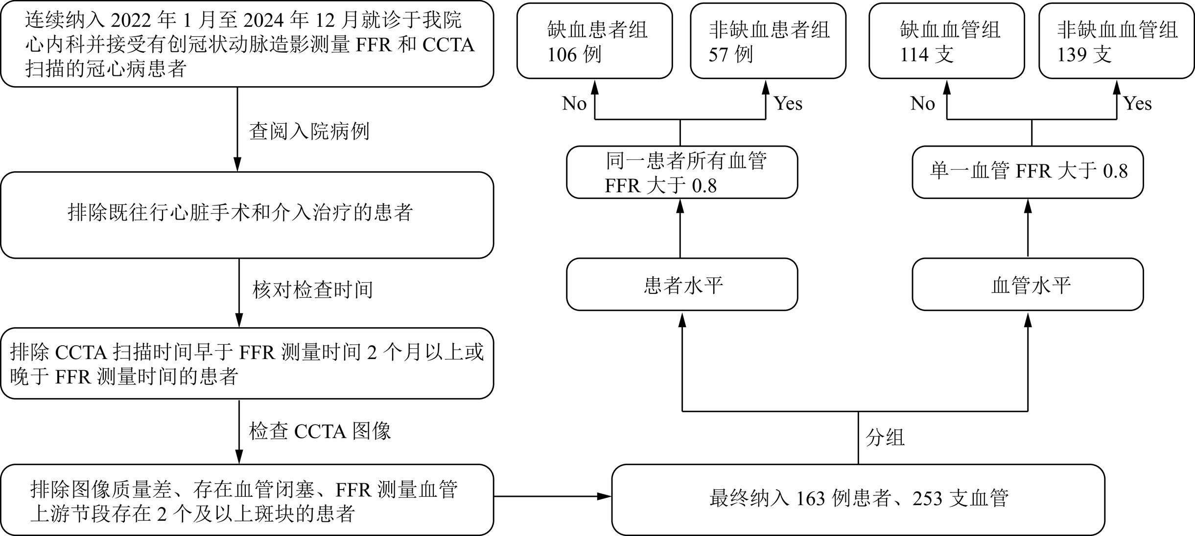
Objective: To compare the differences in coronary computed tomography angiography (CCTA) plaque characteristics between ischemic and non-ischemic groups and to explore qualitative and quantitative plaque features that are valuable for diagnosing myocardial ischemia. This study aimed to apply these indicators in clinical practice to identify patients with potential myocardial ischemia as early as possible. Methods: A retrospective analysis was conducted on patients with coronary heart disease who underwent invasive coronary angiography for fractional flow reserve (FFR) measurement and CCTA scanning in the cardiology department of our hospital between January 2022 and December 2024. General information was analyzed at the patient level, whereas CCTA plaque characteristics were analyzed at the vessel level. Plaque analysis was performed by two radiologists with more than five years of experience in CCTA diagnosis using semi-automatic plaque analysis software blinded to the patient groups. Results: A total of 163 patients were included in the study, with no statistically significant differences in general information between the ischemic and non-ischemic groups. A total of 253 vessels were included, with 114 vessels in the ischemic and 139 in the nonischemic vessel groups. Among the qualitative plaque characteristics, the napkin-ring sign and punctate calcification were more prevalent in the ischemic than in the nonischemic vessel group. No statistically significant differences were observed in positive remodeling and low-density plaque indicators between the two groups. Among the quantitative plaque characteristics, statistically significant differences in plaque length (PL), plaque burden (PB), minimum lumen area (MLA), minimum diameter stenosis (MDS), maximum area stenosis (MAS), and remodeling index (RI) were observed between the two groups. There were no statistically significant differences in plaque volume (PV) or edge irregularity (EI). The areas under the curve (AUCs) for diagnosing myocardial ischemia in the vascular supply areas of the plaques using PL, PB, MLA, MDS, MAS, and RI were 0.672, 0.712, 0.843, 0.830, 0.821, and 0.655, respectively. The AUC for the combined detection was 0.844, which was higher than that for any single indicator. Conclusion: CCTA plaque characteristic analysis has great potential for predicting myocardial ischemia, and the combined use of multiple quantitative plaque indicators provides higher diagnostic efficacy.
| [1] |
国家心血管病中心, 中国心血管健康与疾病报告编写组, 胡盛寿. 中国心血管健康与疾病报告2023概要[J]. 中国循环杂志, 2024, 39(07): 625-660. DOI: 10.3969/j.issn.1000-3614.2024.07.001.
NATIONAL CENTER FOR CARDIOVASCULAR DISEASES, THE WRITING COMMITTEE OF THE REPORT ON CARDIOVASCULAR HEALTH AND DISEASES IN CHINA, HU S S. Report on cardiovascular health and diseases in China 2023: an updated summary[J]. Chinese Circulation Journal, 2024, 39(07): 625-660. DOI: 10.3969/j.issn.1000-3614.2024.07.001.
|
| [2] |
专家组中国冠状动脉血流储备分数测定技术临床路径专家共识. 中国冠状动脉血流储备分数测定技术临床路径专家共识[J]. 中国介入心脏病学杂志, 2019, 27(3): 121-133. DOI: 10.3969/j.issn.1004-8812.2019.03.001.
|
| [3] |
闫昕, 赵建华. 基于CCTA的冠状动脉周围脂肪组织影像组学研究进展[J]. CT理论与应用研究, 2024, 33(4): 531-538. DOI: 10.15953/j.ctta.2023.179.
YAN X, ZHAO JH. Research progress of pericoronary adipose tissue radiomics based on coronary computed tomography angiography[J]. CT Theory and Applications, 2024, 33(4): 531-538. DOI: 10.15953/j.ctta.2023.179.
|
| [4] |
胸痛中心专家委员会, 中华医学会心电生理和起搏分会, 中国医师协会心律学专业委员会, 等. 冠状动脉粥样硬化性心脏病猝死防治专家共识(2024)[J]. 中华心血管病杂志(网络版), 2024, 07(1): 1-18. DOI: 10.3760/cma.j.cn116031.2024.1000177.
|
| [5] |
高扬, 吕滨. 冠状动脉CT血管成像最新临床应用推荐及诊断规范[J]. 中华放射学杂志, 2022, 56(10): 1160-1164. DOI: 10.3760/cma.j.cn112149-20220630-00555.
GAO Y, LV B. The latest clinical application recommendation and diagnostic criteria of coronary CT angiography[J]. Chinese Journal of Radiology, 2022, 56(10): 1160-1164. DOI: 10.3760/cma.j.cn112149-20220630-00555.
|
| [6] |
LEE J M, CHOI G, KOO B, et al. Identification of high-risk plaques destined to cause acute coronary syndrome using coronary computed tomographic angiography and computational fluid dynamics[J]. JACC: Cardiovascular Imaging, 2019, 12(6): 1032-1043. DOI: 10.1016/j.jcmg.2018.01.023.Epub2018Mar14.
|
| [7] |
李正腾, 王敏, 潘冬梅, 等. CCTA斑块特征在冠状动脉管腔狭窄程度进展预测及预后的价值研究[J]. CT理论与应用研究, 2025, 34(1): 23-30. DOI: 10.15953/j.ctta.2024.172.
LI Z T, WANG M, PAN D M, et al. Predictive Value of CCTA Plaque Characteristics for the Progression and Prognosis of Coronary Artery Stenosis[J]. CT Theory and Applications, 2025, 34(1): 23-30. DOI: 10.15953/j.ctta.2024.172.
|
| [8] |
高雪莲, 王瑞, 张宏凯, 等. 冠状动脉CT血管造影影像组学用于冠心病研究进展[J]. 中国医学影像技术, 2024, 40(3): 451-454. DOI: 10.13929/j.issn.1003-3289.2024.03.027.
GAO X L, WANG R, ZHANG H K et al. Research progresses of radiomics based on coronary CT angiography in coronary artery disease[J]. Chinese Journal of Medical Imaging Technology, 2024, 40(3): 451-454. DOI: 10.13929/j.issn.1003-3289.2024.03.027.
|
| [9] |
DESEIVE S, KUPKE M, STRAUB R, et al. Quantified coronary total plaque volume from computed tomography angiography provides superior 10-year risk stratification[J]. European Heart Journal cardiovascular Imaging, 2021, 22(3): 314-321. DOI: 10.1093/ehjci/jeaa228.
|
| [10] |
JIAN Z, YAO G, GUO H, et al. The impact of baseline calcified plaque volume on coronary rapid plaque progression by serial coronary computed tomography angiography in patients with type 2 diabetes[J]. Annals of Medicine, 2023, 55(1): 2196438. DOI: 10.1080/07853890.2023.2196438.
|
| [11] |
盛玉杰, 王询, 王泽静. CT冠状动脉定量在评估冠心病患者心肌缺血诊断中的应用价值[J]. 中国CT和MRI杂志, 2024, 22(5): 103-105. DOI: 10.3969/j.issn.1672-5131.2024.05.033.
SHENG Y J, WANG X, WANG Z J. Application value of coronary CT angiography quantification in the diagnosis of myocardial ischemia in patients with coronary heart disease[J]. Chinese Journal of CT and MRI, 2024, 22(5): 103-105. DOI: 10.3969/j.issn.1672-5131.2024.05.033.
|
| [12] |
孙俊, 夏花, 江时忠. 冠状动脉CT血管成像定量评估冠心病心肌缺血的效能[J]. 浙江医学, 2022, 44(6): 637-640, 645. DOI: 10.12056/j.issn.1006-2785.2022.44.6.2021-2378.
SUN J, XIA H, JIANG S Z. Value of coronary CT angiography in quantitative assessment of myocardial ischemia in patients with coronary heart disease[J]. Zhejiang Medical Journal 2022, 44(6): 637-640, 645. DOI: 10.12056/j.issn.1006-2785.2022.44.6.2021-2378. (in Chinese).
|
| [13] |
CHUNG C J, JEONG S Y, JEONG J H, et al. Comparison of prophylactic effect of topical alchemilla vulgaris in glycerine versus that of dexamethasone on postoperative sore throat after tracheal intubation using a double-lumen endobronchial tube: a randomized controlled study[J]. Anesthesia and Pain Medicine, 2021, 16(2): 163-170. DOI: 10.17085/apm.20082.
|
| [14] |
高艳, 顾慧, 杨世锋, 等. 基于冠状动脉CT血管成像的斑块定量分析及其与心肌缺血损伤的相关性研究[J]. 中华放射学杂志, 2020, 54(2): 7. DOI: 10.3760/cma.j.issn.1005-1201.2020.02.008.
GAO Y, GU H, YANG S, et al. Correlation study of coronary plaque quantitative analysis and myocardial ischemic injury based on coronary CT angiography[J]. Chinese Journal Radiology, 2020, 54(2): 7. DOI: 10.3760/cma.j.issn.1005-1201.2020.02.008.
|
| [15] |
LONG Y, GUO R, JIN K, et al. Analysis of the perivascular fat attenuation index and quantitative plaque parameters in relation to hemodynamically impaired myocardial ischemia[J]. The International Journal of Cardiovascular Imaging, 2024, 40(7): 1455-1463. DOI: 10.1007/s10554-024-03122-x.
|
| [16] |
BAR S, MAANIITTY T, KIATKITTIKUL P, et al. Incremental prognostic value of artificial intelligence-based automated plaque characterisation on top of ischemia evaluation by CCTA and CCTA/PET[J]. European Heart Journal Cardiovascular Imaging, 2024, 25(Sup1): 1. DOI: 10.1093/ehjci/jeae142.036.
|
| [17] |
SAITO Y, KOBAYASHI Y, FUJII K, et al. CVIT 2023 clinical expert consensus document on intravascular ultrasound[J]. Cardiovascular Intervention and Therapeutics, 2024, 39(1): 1-14. DOI: 10.1007/s12928-023-00957-4.
|
| [18] |
LIN A, MANRAL N, MCELHINNEY P, et al. Deep learning-enabled coronary CT angiography for plaque and stenosis quantification and cardiac risk prediction: an international multicentre study[J]. Lancet Digit Health, 2022, 4(4): e256-e265. DOI: 10.1016/S2589-7500(22)00022-X.
|
| [19] |
KAWASAKI T, KIDOH M, KIDO T et al. Evaluation of significant coronary artery disease based on CT fractional flow reserve and plaque characteristics using random forest analysis in machine learning[J]. Academic Radiology, 2020, 27(12): 1700-1708. DOI: 10.1016/j.acra.2019.12.013.
|
| [20] |
朱娜君, 方欣欣, 尹伊君, 等. 心绞痛患者斑块进展危险因素与冠状动脉CT血管成像指标的关系研究[J]. CT理论与应用研究, 2023, 32(2): 217-222. DOI: 10.15953/j.ctta.2022.219.
ZHU N J, FANG X X, YIN Y J et al. Risk factors of plaque progression in patients with angina pectoris and their relationships with coronary CT angiography[J]. CT Theory and Applications, 2023, 32(2): 217-222. DOI: 10.15953/j.ctta.2022.219.
|
| [1] | LIU Xiaoyan, BAO Zhongying, DUAN Shuhong, ZHANG Jie, ZHANG Mingxia, SUN Ying, LI Ling, WANG Rengui. Clinical Characteristics and Imaging Features of COVID-19 at Initial Diagnosis in Fever Clinic[J]. CT Theory and Applications, 2023, 32(5): 636-644. DOI: 10.15953/j.ctta.2023.149 |
| [2] | ZHANG Mingxia, LI Ling, SUN Ying, GUO Jia, DU Changyue, LI Xingpeng, ZHANG Yan, HAO Qi, DUAN Shuhong, LIU Xiaoyan, SUN Lei, HUO Meng, ZHANG Chunyan, WANG Rengui. Comparative Analysis of Clinical and Computed Tomography Imaging Features of COVID-19 with Different Disease Courses[J]. CT Theory and Applications, 2023, 32(3): 380-386. DOI: 10.15953/j.ctta.2023.021 |
| [3] | CHE Hongwei, ZHANG Xiaoqin, CHAI Jun, SUN Dejun. Clinical Manifestations and CT Imaging Analysis of Corona Virus Disease 2019[J]. CT Theory and Applications, 2021, 30(4): 525-532. DOI: 10.15953/j.1004-4140.2021.30.04.14 |
| [4] | ZHANG Hecheng, CHU Yan, LIU Jing, LI Xiaozhen, ZHAO Tianzuo. The Clinical Features and CT Manifestations of the Novel Coronavirus Pneumonia COVID-19[J]. CT Theory and Applications, 2020, 29(5): 559-565. DOI: 10.15953/j.1004-4140.2020.29.05.06 |
| [5] | WANG Zengkui, ZHANG Zhaofu, PANG Jun, WEI Xiaohua, PANG Hongyan, GAO Dongwei. The Clinical Subtypes of Corona Virus Disease 2019 Correspond to CT Findings and the Value of Artificial Intelligence[J]. CT Theory and Applications, 2020, 29(5): 534-542. DOI: 10.15953/j.1004-4140.2020.29.05.03 |
| [6] | WANG Gang, XIE Haofeng, ZHENG Xiaolin, FANG Xuewen, YU Fenfen, YUAN Huanchu, DU Heqin, ZOU Yujian. The Initial Study of Clinical and CT Diagnostic Characteristics about Corona Virus Disease 2019: Case Review in Dongguan[J]. CT Theory and Applications, 2020, 29(4): 407-415. DOI: 10.15953/j.1004-4140.2020.29.04.03 |
| [7] | YIN Ke, QIU Taichun, WANG Qiong, WU Jianlin. Clinical and CT Manifestations of Corona Virus Disease 2019[J]. CT Theory and Applications, 2020, 29(3): 281-287. DOI: 10.15953/j.1004-4140.2020.29.03.03 |
| [8] | JU Min-hao, GUO Yue-feng, WANG Yan-yan, ZHANG Xiao-lu. Analysis of Clinical Manifestations and CT and MRI Findings of Venous Cerebral Embolism[J]. CT Theory and Applications, 2016, 25(5): 619-624. DOI: 10.15953/j.1004-4140.2016.25.05.15 |
| [9] | WU De-hong, CHEN Shao-bo, MU Hua-guo, GONG Xiao-hong, FU Chuan-ming, CHEN Wen. CT and MRI Manifestations of Autoimmune Pancreatitis[J]. CT Theory and Applications, 2015, 24(4): 611-619. DOI: 10.15953/j.1004-4140.2015.24.04.16 |
| [10] | HUANG Shao-quan, YE Jun, CHEN Xue-lian, ZOU Shi-lin, KUANG Yong-cai, LIU Yong-bin. CT Analysis of Degenerative Lesion in Lumbar Zygapophysis[J]. CT Theory and Applications, 2011, 20(1): 115-122. |
Supported by: Beijing Renhe Information Technology Co. Ltd 

 DownLoad: