ISSN 1004-4140
CN 11-3017/P
| Citation: |
CHEN Z G, WEI N N, LI Z F, et al. Methods Research on Correction for Intracranial Metal Artifacts Based on Prior Image Utilization[J]. CT Theory and Applications, 2025, 34(1): 141-148. DOI: 10.15953/j.ctta.2024.002. (in Chinese).

|
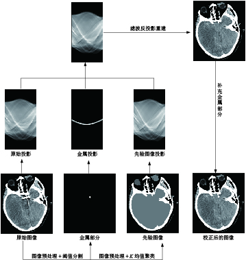
Objective: A metal artifact correction algorithm based on prior image is proposed to suppress the effect of intracranial metal artifacts on postoperative review. Method: The computed tomography (CT) image compensating for the intracranial metal artifact is obtained by adaptive filtering, threshold segmentation, and K mean clustering. In projecting the trajectory of metallic matter, the interpolation correction of the original image’s projection data is obtained using the prior image as a reference, and the image is fused with the metal-containing material only image to produce the final corrected image. To verify the efficacy of this algorithm in suppressing CT metal artifacts, artifact correction was performed using CT images of simulated isolated metal artifacts and intracranial metal artifacts. Results: Compared with linear interpolation and the ADN algorithms, this algorithm achieves the highest structure similarity and peak signal-to-noise ratio and lowest root mean square error in corrected images. Two senior doctors subjectively scored this algorithm higher than the linear interpolation and ADN algorithms for removing CT metal artifacts. Conclusion: This algorithm effectively suppresses metal artifacts while preserving image edge information.
| [1] |
DING Y H, GHOZY S, DAI D, et al. Rabbit elastase aneurysm model mimics the recurrence rate of human intracranial aneurysms following platinum coil embolization[J]. American Journal of Neuroradiology. 2022, 43(5): 741-747.
|
| [2] |
刘远来, 孙异春, 何永超, 等. Solitaire支架与lvis支架辅助弹簧圈栓塞对颅内宽颈动脉瘤的疗效对比[J]. 南方医科大学学报, 2020, 40(3): 423-426. DOI: 10.12122/j.issn.1673-4254.2020.03.23.
LIU Y L, SUN Y C, HE Y C, et al. Treatment efficacy of Solitaire stent- and Lvis stent-assisted coil embolization for intracranial wide-neck carotid aneurysm[J]. Journal of Southern Medical University, 2020, 40(3): 423-426. DOI:10.12122/j.issn.1673-4254.2020.03.23. (in Chinese).
|
| [3] |
DAI W, LING H, SUN Y, et al. The comparison of neuronavigation combined with CT three-dimensional angiography vs. CT angiography in the guidance of clipping treatment in distal intracranial aneurysm surgery: A retrospective clinical study[J]. Annals of Translational Medicine, 2022, 10(10): 572-578.
|
| [4] |
KANANI A, YAZDI M, OWRANGI A M, et al. Metal artifact reduction in cervix brachytherapy with titanium applicators using dual-energy CT through virtual monoenergetic images and an iterative algorithm: A phantom study[J]. Brachytherapy, 2022, 21(6): 933-942.
|
| [5] |
陈宗桂, 陆思璇, 管海辰, 等. IMAR技术在去除腰椎内固定金属伪影中的应用价值[J]. 中国CT和MRI杂志, 2023, 21(11): 167-169. DOI: 10.3969/j.issn.1672-5131.2023.11.051.
CHEN Z G, LU S X, GUAN H C, et al. Value of IMAR Iterative algorithm in removing metal artifacts of lumbarInternal fixation[J]. Chinese Journal of CT and MRI, 2023, 21(11): 167-169. DOI:10.3969/j.issn.1672-5131.2023.11.051. (in Chinese).
|
| [6] |
SCHWARZ G M, HUBER S, WASSIPAUL C, et al. Influence of scan parameters of single and dual-energy CT protocols in combination with metal artifact suppression algorithms for THA: An EX Vivo study[J]. Journal of Bone and Joint Surgery-american Volume, 2023, 105(8): 620-629.
|
| [7] |
于佳弘, 张昆鹏, 靳爽, 等. 弦图插值结合UNIT网络图像转换的CT金属伪影校正[J]. 南方医科大学学报, 2023, 43(7): 1214-1223. DOI: 10.12122/j.issn.1673-4254.2023.07.18.
YU J H, ZHANG K P, JIN S, et al. CT metal artifact correction using chord graph interpolation combined with UNIT network image conversion[J]. Journal of Southern Medical University, 2023, 43(7): 1214-1223. DOI: 10.12122/j.issn.1673-4254.2023.07.18. (in Chinese).
|
| [8] |
ZHANG Z, YANG M, XU L, et al. An innovative metal artifact reduction algorithm based on Res-U-Net GANs[J]. Current Medical Imaging, 2023, 19(13): 1549-1560.
|
| [9] |
YOON H, LEE K Y, BECHWATI I. CLIMAR: Classified linear interpolation based metal artifact reduction for severe metal artifact reduction in X-ray CT imaging[J]. Physics in Medicine&Biology, 2021, 66(7): 1-13.
|
| [10] |
齐宏亮, 凌庆, 李翰威, 等. 基于先验图像的CT图像金属伪影校正算法[J]. 北京生物医学工程, 2023, 42(4): 384-389, 413. DOI: 10.3969/j.issn.1002-3208.2023.04.008.
QI H L, LING Q, LI H W, et al. Metal artifact correction algorithm for CT images based on prior images[J]. Befjing Blomedical Engineering, 2023, 42(4): 384-389, 413. DOI: 10.3969/j.issn.1002-3208.2023.04.008. (in Chinese).
|
| [11] |
SCHMIDT A M A, GRUNZ J P, PETRITSCH B, et al. Combination of iterative metal artifact reduction and virtual monoenergetic reconstruction using split-filter dual-energy CT in patients with dental artifact on head and neck CT[J]. American Journal of Roentgenology, 2022, 218(4): 716-727.
|
| [12] |
TRABZONLU T A, TERRAZAS M, MOZAFFARY A, et al. Application of iterative metal artifact reduction algorithm to CT urography for patients with hip prostheses[J]. American Journal of Roentgenology, 2020, 214(1): 137-143.
|
| [13] |
JACOBS F, SUNDERMANN E, de Sutter B, et al. A fast algorithm to calculate the exact radiological path through a pixel or voxel space[J]. Journal of Computing and Information Technology, 1998, 6(1): 89-94.
|
| [14] |
LIAO H F, LIN W A, ZHOU S K, et al. ADN: Artifact disentanglementnetwork for unsupervised metal artifact reduction[J]. IEEE TransMed Imaging, 2020, 39(3): 634-643. DOI: 10.1109/TMI.2019.2933425.
|
| [15] |
王军. 基于投影数据修正的CT金属伪影消除方法研究[D]. 南京: 东南大学, 2013. DOI: 10.7666/d.Y2438396.
WANG J. Research on CT metal artifact elimination method based on projection data correction[D]. Nanjing: Southeast University, 2013. DOI:10.7666/d.Y2438396.(in Chinese).
|
| [16] |
SARA U, AKTER M, UDDIN M S. Image quality assessment through FSIM, SSIM, MSE and PSNR-A comparative study[J]. Journal of Computer and Communications, 2019, 7(3): 8-18. DOI: 10.4236/jcc.2019.73002.
|
| [17] |
MATTHIEU B, LOTHAR S. Metal artifact reduction in CT using tissue-class modeling and adaptive prefiltering[J]. Medical Physics, 2006, 33(8): 2852-2859. DOI: 10.1118/1.2218062.
|
| [18] |
郑艳梅, 岳向江, 李翠, 等. 基于均方误差的变形图像的质量评价[J]. 测控技术, 2018, 37(8): 117-120. DOI: 10.19708/j.ckjs.2018.08.027.
ZHENG Y M, YUE X J, LI C, et al. Quality evaluation of deformed images based on mean square error[J]. Measurement and Control Technology, 2018, 37(8): 117-120. DOI: 10.19708/j.ckjs.2018.08.027. (in Chinese).
|
| [19] |
ANHAUS J A, KILLERMANN P, SEDLMAIR M, et al. Nonlinearly scaled prior image-controlled frequency split for high-frequency metal artifact reduction in computed tomography[J]. Medical Physics, 2022, 49(9): 5870-5885.
|
| [20] |
GONDIM T P, MEYER J B, BAUMANN C, et al. Total hip prosthesis CT with single-energy projection based metallic artifact reduction: Impact on the visualization of specific periprosthetic soft tissue structures[J]. Skeletal Radiology, 2014, 43(9): 1237-1246. DOI: 10.1007/s00256-014-1923-5.
|
| [1] | LIU Yuxin, WEI Jiaotong, ZHAO Xiaojie, CHEN Ping, PAN Jinxiao. A Correction Method for Hardening Artifacts in CT Images Based on Integral Invariance[J]. CT Theory and Applications, 2025, 34(4): 571-579. DOI: 10.15953/j.ctta.2025.064 |
| [2] | SU Danyang, HOU Ping, ZHANG Haoran, MA Yuanbo, LIU Jinlong, YANG Xiaopeng. Research Progress on Reducing Metal Artifacts in CT Imaging[J]. CT Theory and Applications, 2025, 34(3): 499-505. DOI: 10.15953/j.ctta.2024.023 |
| [3] | LIU Xiangcheng, HUANG Xiaoying, WEI Pengyue, WANG Pengchao, GUO Fangkai, BAO Yunfeng. Clinical Research Progress on Deep Learning for Metal Artifact Reduction in CT Images[J]. CT Theory and Applications. DOI: 10.15953/j.ctta.2025.222 |
| [4] | WEI Pengyue, HUANG Xiaoying, WU Tong, GUO Fangkai, GUAN Jing, BAO Yunfeng. Effect of denture materials on CT image artifacts: phantom experiment[J]. CT Theory and Applications. DOI: 10.15953/j.ctta.2024.339 |
| [5] | LIU Bicheng, YI Xi, ZONG Chunguang, XU Yanwei, LI Liang. Ring-artifact Correction Method for Large-size Object CT Images Based on Gradient Featured Cluster Analysis[J]. CT Theory and Applications, 2024, 33(6): 781-789. DOI: 10.15953/j.ctta.2024.153 |
| [6] | ZUO Shunji, FENG Peng, HUANG Pan, YAN Shenghao, HE Peng, WEI Biao. Metal Artifact Reduction Algorithm for CT Images of Rock and Mineral Samples Based on Dual-domain Adaptive Network[J]. CT Theory and Applications, 2022, 31(6): 783-792. DOI: 10.15953/j.ctta.2022.041 |
| [7] | ZHAO Qingqing, SUN Xiaoli, LI Xi, WANG Yanwen, LIU Weina, DUAN Yongli, WANG Rengui. Utility of Spectral CT Monochromatic Imaging with Metal Artifacts Reduction (MAR) for the Reduction of Metal Artifacts of Embolization Coil Implants[J]. CT Theory and Applications, 2019, 28(5): 529-539. DOI: 10.15953/j.1004-4140.2019.28.05.02 |
| [8] | ZHANG Xue-song, ZHAO Bo-shan. Cupping Artifacts Calibration in CT Image Based on Radon Transform[J]. CT Theory and Applications, 2016, 25(5): 539-546. DOI: 10.15953/j.1004-4140.2016.25.05.05 |
| [9] | CHAI Jia-bin, LI Gong-ping, WEI Cun-feng, WEI Long, YU Yang, WANG Qian, LIU Bao-dong. Metal Artifacts Correction Method Research about Liquid Metal Angiography[J]. CT Theory and Applications, 2016, 25(2): 149-157. DOI: 10.15953/j.1004-4140.2016.25.02.04 |
| [10] | LI Qing-liang, YAN Bin, SUN Hong-sheng, LI Lei, ZHANG Feng. Review of Metal Artifacts Correction Methods on X-ray Computed Tomography[J]. CT Theory and Applications, 2011, 20(1): 131-140. |
Supported by: Beijing Renhe Information Technology Co. Ltd 


 DownLoad: