ISSN 1004-4140
CN 11-3017/P
| Citation: |
WANG R, YANG J, CHEN Y, et al. Correlation Between MRI Changes and Bone Mineral Density in Patients with Knee Osteoarthritis[J]. CT Theory and Applications, 2024, 33(5): 601-608. DOI: 10.15953/j.ctta.2024.024. (in Chinese).

|
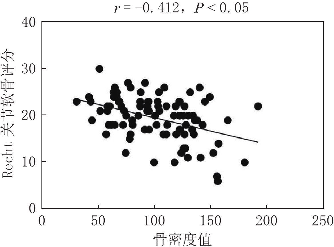
Objective: To determine the relationship between cartilage injury, bone marrow edema, and total bone mineral density in patients with knee osteoarthritis (KOA). Methods: Ninety patients with KOA admitted to the Sichuan Province Orthopedic Hospital between March 2023 and October 2023 were selected, all of whom underwent knee joint MRI and chest QCT examinations. The patients were divided into three groups according to the QTC results: normal bone density, reduced bone mass, and osteoporosis. Patient information, such as sex, age, body mass index (BMI), and course of illness, was also obtained. MRIs of the knee joints were scored separately according to the Recht grading and Whole-organ Magnetic Resonance Imaging Score (WORMS). The bone mineral density (BMD) of the patients was calculated and the BMD values were statistically analyzed using the MR score. Results: No statistically significant differences were observed in sex, BMI, or disease duration among the three groups; however, a statistically significant difference was observed in age. The Recht and WORMS scores among the three groups showed statistical significance between the normal bone density group and the reduced bone mass and osteoporosis groups; however, no statistical significance was observed between the reduced bone mass and osteoporosis groups. KOA cartilage injury was negatively correlated with bone density with a correlation coefficient of r=−0.412, while KOA bone marrow edema was negatively correlated with bone density with a correlation coefficient of r=−0.254. Conclusion: The total bone density in patients with KOA is related to knee joint cartilage injury and bone marrow edema. A decrease in total bone density may indicate the severity of knee cartilage injury or bone marrow edema.
| [1] |
王斌, 邢丹, 董圣杰, 等. 中国膝骨关节炎流行病学和疾病负担的系统评价[J]. 中国循证医学杂志, 2018, 18(2): 134−142.
WANG B, XING D, DONG S J, et al. Prevalence and disease burden of knee osteoarthritis in China: A systematic review[J]. Chinese Journal of Evidence-based Medicine, 2018, 18(2): 134−142. (in Chinese).
|
| [2] |
HARRELL C R, MARKOVIC B S, FELLABAUM C, et al. Mesenchymal stem cell-based therapy of osteoarthritis: Current knowledge and future perspectives[J]. Biomedicine Pharmacotherapy, 2019, 109: 2318−2326. DOI: 10.1016/j.biopha.2018.11.099.
|
| [3] |
MESSENT E A, WARD R J, TONKIN C J, et al. Differences in trabecular structure between knees with and without osteoarthritis quantified by macro and standard radiography, respectively[J]. Osteoarthritis Cartilage, 2006, 14(12): 1302−1305. DOI: 10.1016/j.joca.2006.07.012.
|
| [4] |
BUCKLAND-WRIGHT C. Subchondral bone changes in hand and knee osteoarthritis detected by radiography[J]. Osteoarthritis Cartilage, 2004, 12(S)A: S10-S19.
|
| [5] |
DEDRICK D K, GOLDSTEIN S A, BRANDT K D, et al. A longitudinal study of subchondral plate and trabecular bone in cruciate-deficient dogs with osteoarthritis followed up for 54 months[J]. Arthritis Rheumatic, 1993, 36(10): 1460−1467. DOI: 10.1002/art.1780361019.
|
| [6] |
张程, 吴忠书, 李子祺, 等. 膝骨关节炎与骨质疏松症的相关性研究进展[J]. 中国骨质疏松杂志, 2021, 27(4): 618−624. DOI: 10.3969/j.issn.1006-7108.2021.04.028.
ZHANG C, WU Z S, LI Z Q, et al. Research progress on the correlation between knee osteoarthritis and osteoporosis[J]. Chinese Journal of Osteoporosis, 2021, 27(4): 618−624. DOI: 10.3969/j.issn.1006-7108.2021.04.028. (in Chinese).
|
| [7] |
BENNELL K L, CREABY M W, WRIGLEY T V, et al. Bone marrow lesions are related to dynamic knee loading in medial knee osteoarthritis[J]. Annals of the Rheumatic Diseases, 2010, 69(6): 1151−1154. DOI: 10.1136/ard.2009.118182.
|
| [8] |
王勇朋, 阳琰, 何生生, 等. 低剂量胸部CT与QCT椎体骨密度测量一站式扫描可行性研究[J]. 放射学实践, 2018, 33(11): 1194−1197.
WANG Y P, YANG Y, HE S S, et al. Low-dose chest CT and QCT “one-stop-shop” scan technology: A feasibility study[J]. Radiologic Practice, 2018, 33(11): 1194−1197. (in Chinese).
|
| [9] |
中华医学会骨科学分会关节外科学组. 骨关节炎诊疗指南(2018年版)[J]. 中华骨科杂志, 2018, 38(12): 705−715. DOI: 10.3760/cma.j.issn.0253-2352.2018.12.001.
Osteoporosis Group of Chinese Orthopaedic Associationg. Chinese guideline for diagnosis and management of osteoarthritis (2018 edition)[J]. Chinese Journal of Orthopaedics, 2018, 38(12): 705−715. DOI: 10.3760/cma.j.issn.0253-2352.2018.12.001. (in Chinese).
|
| [10] |
RECHT M P, KRAMER J, MARCELIS S, et al. Abnormalities of articular cartilage in the knee: Analysis of available MR techniques[J]. Radiology, 1993, 187(2): 473−478. DOI: 10.1148/radiology.187.2.8475293.
|
| [11] |
PETERFY C G, GUERMAZI A, ZAIM S, et al. Whole-organ magnetic resonance imaging score (WORMS) of the knee in osteoarthritis[J]. Osteoarthritis Cartilage, 2004, 12(3): 177−190. DOI: 10.1016/j.joca.2003.11.003.
|
| [12] |
李凯, 陈捷, 赵林芬, 等. 中国人群定量CT(QCT)脊柱骨密度正常参考值的建立和骨质疏松症QCT诊断标准的验证[J]. 中国骨质疏松杂志, 2019, 25(9): 1257−1262. DOI: 10.3969/j.issn.1006-7108.2019.09.011.
LI K, CHEN J, ZHAO L F, et al. The establishment of QCT spinal vBMD re ference database and the validation of the diagnosis criteria of oste oporosis with QCT for Chinese[J]. Chinese Journal of Osteoporosis, 2019, 25(9): 1257−1262. DOI: 10.3969/j.issn.1006-7108.2019.09.011. (in Chinese).
|
| [13] |
舒意, 杨沛, 廖紫祾, 等. 一站式低剂量扫描在胸部及腰椎QCT体检人群中的应用[J]. CT理论与应用研究, 2022, 31(2): 244−250. DOI: 10.15953/j.1004-4140.2022.31.02.12.
SHU Y, YANG P, LIAO Z L, et al. To explore the low-dose CT and QCT “one-stop-shop” scan technology for physical examination crowd[J]. CT Theory and Applications, 2022, 31(2): 244−250. DOI: 10.15953/j.1004-4140.2022.31.02.12. (in Chinese).
|
| [14] |
赵敏, 刘鸿雁, 王国华, 等. 膝关节骨关节炎半月板损伤程度与关节软骨T1 rho、T2 mapping相关性研究[J]. 中国临床医学影像杂志, 2019, 30(11): 812−816.
ZHAO M, LIU H Y, WANG G H, et al. The correlation between the degree of meniscus injury of knee osteoarthritis and T1 rho, T2 mapping of the articular cartilage[J]. Journal of China Clinic Medical Imaging, 2019, 30(11): 812−816. (in Chinese).
|
| [15] |
王佳, 朱吉云, 茅博伟, 等. 膝关节骨性关节炎软骨损伤的MRI形态评分及T2值变化与临床表现的相关性[J]. 影像研究与医学应用, 2021, 5(21): 26−27. DOI: 10.3969/j.issn.2096-3807.2021.21.012.
WANG J, ZHU J Y, MAO B W, et al. MRI morphological scores of cartilage injury in knee joint osteoarthritis and correlation between changes of T2 value and clinical features[J]. Journal of Imaging Research and Medical, 2021, 5(21): 26−27. DOI: 10.3969/j.issn.2096-3807.2021.21.012. (in Chinese).
|
| [16] |
刘艳平, 廖忠剑, 李正南. 磁共振成像检查在评估膝关节退行性骨关节炎患者关节损伤程度中的价值[J]. 中国当代医药, 2021, 28(34): 158−161. DOI: 10.3969/j.issn.1674-4721.2021.34.043.
LIU Y P, LIAO Z J, LI Z N. The value of magnetic resonance imaging in assessing the degree of joint damage in patients with knee degenerative osteoarthritis[J]. China Modern Medicine, 2021, 28(34): 158−161. DOI: 10.3969/j.issn.1674-4721.2021.34.043. (in Chinese).
|
| [17] |
韩雪莉. 迭代模型重建技术在低剂量胸部CT联合腰椎QCT扫描中的研究[D]. 郑州: 郑州大学, 2018.
HAN X L. The study of iterative model reconstruction technique on the low dose chest CT combined with lumbar vertebrae quantitative CT[D]. Zhengzhou: Zhengzhou University, 2018. (in Chinese).
|
| [18] |
LEE J Y, HARVEY W F, PRICE L L, et al. Relationship of bone mineral density to progression of knee osteoarthritis[J]. Arthritis Rheumatic, 2013, 65(6): 1541−1546. DOI: 10.1002/art.37926.
|
| [19] |
BOBINAC D, SPANJOL J, ZORICIC S, et al. Changes in articular cartilage and subchondral bone histomorphometry in osteoarthritic knee joints in humans[J]. Bone, 2003, 32(3): 284−290. DOI: 10.1016/S8756-3282(02)00982-1.
|
| [20] |
KAMIBAYASHI L, WYSS U P, COOKE T D, et al. Trabecular microstructure in the medial condyle of the proximal tibia of patients with knee osteoarthritis[J]. Bone, 1995, 17(1): 27−35. DOI: 10.1016/8756-3282(95)00137-3.
|
| [21] |
LI B, ASPDEN R M. Composition and mechanical properties of cancellous bone from the femoral head of patients with osteoporosis or osteoarthritis[J]. Journal of Bone and Mineral Research, 1997, 12(4): 641−651. DOI: 10.1359/jbmr.1997.12.4.641.
|
| [22] |
TOKGÖZ M A, ATIK O Ş, ESENDAĞLI G, et al. Is it possible that the pathogenesis of osteoarthritis could start with subchondral trabecular bone loss like osteoporosis?[J]. Eklem Hastalik Cerrahisi, 2018, 29(3): 152−158. DOI: 10.5606/ehc.2018.007.
|
| [23] |
MARCUCCI G, BRANDI M L. Rare causes of osteoporosis[J]. Clinical Cases in Mineral and Bone Metabolism, 2015, 12(2): 151−156.
|
| [24] |
RYD L, BRITTBERG M, ERIKSSON K, et al. Pre-osteoarthritis: Definition and diagnosis of an elusive clinical entity[J]. Cartilage, 2015, 6(3): 156−165. DOI: 10.1177/1947603515586048.
|
| [25] |
WEN L, SHIN M H, KANG J H, et al. The relationships between bone mineral density and radiographic features of hand or knee osteoarthritis in older adults: Data from the Dong-gu study[J]. Rheumatology (Oxford), 2016, 55(3): 495−503.
|
| [26] |
ROEMER F W, NEOGI T, NEVITT M C, et al. Subchondral bone marrow lesions are highly associated with, and predict subchondral bone attrition longitudinally: The MOST study[J]. Osteoarthritis Cartilage, 2010, 18(1): 47−53. DOI: 10.1016/j.joca.2009.08.018.
|
| [27] |
DORE D, MARTENS A, QUINN S, et al. Bone marrow lesions predict site-specific cartilage defect development and volume loss: A prospective study in older adults[J]. Arthritis Research Therapy, 2010, 12(6): R222. DOI: 10.1186/ar3209.
|
| [28] |
de SÁ G A, DOS S A, NOGUEIRA J M, et al. Angiotensin II triggers knee joint lesions in experimental osteoarthritis[J]. Bone, 2021, 145: 115842. DOI: 10.1016/j.bone.2021.115842.
|
| [29] |
HEILMEIER U, WAMBA J M, JOSEPH G B, et al. Baseline knee joint effusion and medial femoral bone marrow edema, in addition to MRI-based T2 relaxation time and texture measurements of knee cartilage, can help predict incident total knee arthroplasty 4~7 years later: Data from the Osteoarthritis Initiative[J]. Skeletal Radiology, 2019, 48(1): 89−101. DOI: 10.1007/s00256-018-2995-4.
|
| [30] |
曾国庆, 董铿, 黄建军. 膝骨关节炎与骨质疏松症的相关性分析[J]. 中国卫生标准管理, 2023, 14(18): 94−97. DOI: 10.3969/j.issn.1674-9316.2023.18.022.
ZENG G Q, DONG K, HUANG J J. Analysis of the correlation between knee osteoarthritis and osteoporosis[J]. China Health Standard Management, 2023, 14(18): 94−97. DOI: 10.3969/j.issn.1674-9316.2023.18.022. (in Chinese).
|
| [31] |
周自明, 常时新, 田芳, 等. 膝骨关节炎软骨下骨髓水肿样及囊样病变与软骨病损的相关性研究[J]. 中国医学计算机成像杂志, 2012, 18(3): 243−248. DOI: 10.3969/j.issn.1006-5741.2012.03.012.
ZHOU Z M, CHANG S X, TIAN F, et al. Relationship between subchondral bone marrow edema-like or cyst-like lesions and cartilage loss in patients with knee osteoarthritis[J]. Chinese Computed Medical Imaging, 2012, 18(3): 243−248. DOI: 10.3969/j.issn.1006-5741.2012.03.012. (in Chinese).
|
| [32] |
何勇, 张乾, 高华利, 等. 类风湿关节炎膝关节骨髓水肿病变的影像学研究[J]. 中国矫形外科杂志, 2019, 27(5): 421−425.
HE Y, ZHANG Q, GAO H L, et al. Bone marrow edema in rheumatoid arthritis knee: A study based on MRI[J]. Orthopedic Journal of China, 2019, 27(5): 421−425. (in Chinese).
|
| [33] |
肖龙文, 桑志成. 重度膝骨关节炎患者骨髓水肿与骨质疏松的相关性研究[J]. 中国骨伤, 2023, 36(4): 371−375.
XIAO L W, SANG Z C. Study on correlation between bone marrow edema and osteoporosis in patients with severe knee osteoarthritis[J]. China Journal of Orthopaedics and Traumatology, 2023, 36(4): 371−375. (in Chinese).
|
| [34] |
郭子瑊, 毛兴佳, 高英杰, 等. 膝骨关节炎与膝关节周围骨密度的相关性研究进展[J]. 中华关节外科杂志(电子版), 2021, 15(5): 586−595.
GUO Z J, MAO X J, GAO Y J, et al. Research progress on relationship between knee osteoarthritis and bone mineral density around knee joint[J]. Chinese Journal of Joint Surgery (Electronic Edition), 2021, 15(5): 586−595. (in Chinese).
|
| [35] |
程晓光, 王亮, 曾强, 等. 中国定量CT(QCT)骨质疏松症诊断指南(2018)[J]. 中国骨质疏松杂志, 2019, 25(6): 733−737.
CHENG X G, WANG L, ZENG Q, et al. The China guideline for the diagnosis criteria of osteoporosis with quantitative computed tomography (QCT) (2018)[J]. Chinese Journal of Osteoporosis, 2019, 25(6): 733−737. (in Chinese).
|
| [36] |
ENGELKE K, ADAMS J E, ARMBRECHT G, et al. Clinical use of quantitative computed tomography and peripheral quantitative computed tomography in the management of osteoporosis in adults: The 2007 ISCD official positions[J]. Journal of Clinical Densitometry, 2008, 11(1): 123−162. DOI: 10.1016/j.jocd.2007.12.010.
|
| 1. |
许鹏君,许晨思,孙钢,张翼,赵苏莉,冯亚斌,刘锴. 膝骨性关节炎关节状态与西安大略和麦克马斯特大学骨关节炎指数评分水平的定量CT研究. 创伤与急危重病医学. 2025(02): 145-147+160 .

|
Supported by: Beijing Renhe Information Technology Co. Ltd 


 DownLoad: