ISSN 1004-4140
CN 11-3017/P
| Citation: |
LUO C, REN Q, LIU J Q, et al. Development of motion artifact correction solutions for the cone-beam CT images during pancreatic cancer image-guided radiotherapy[J]. CT Theory and Applications, 2022, 31(6): 761-771. DOI: 10.15953/j.ctta.2022.066. (in Chinese).

|
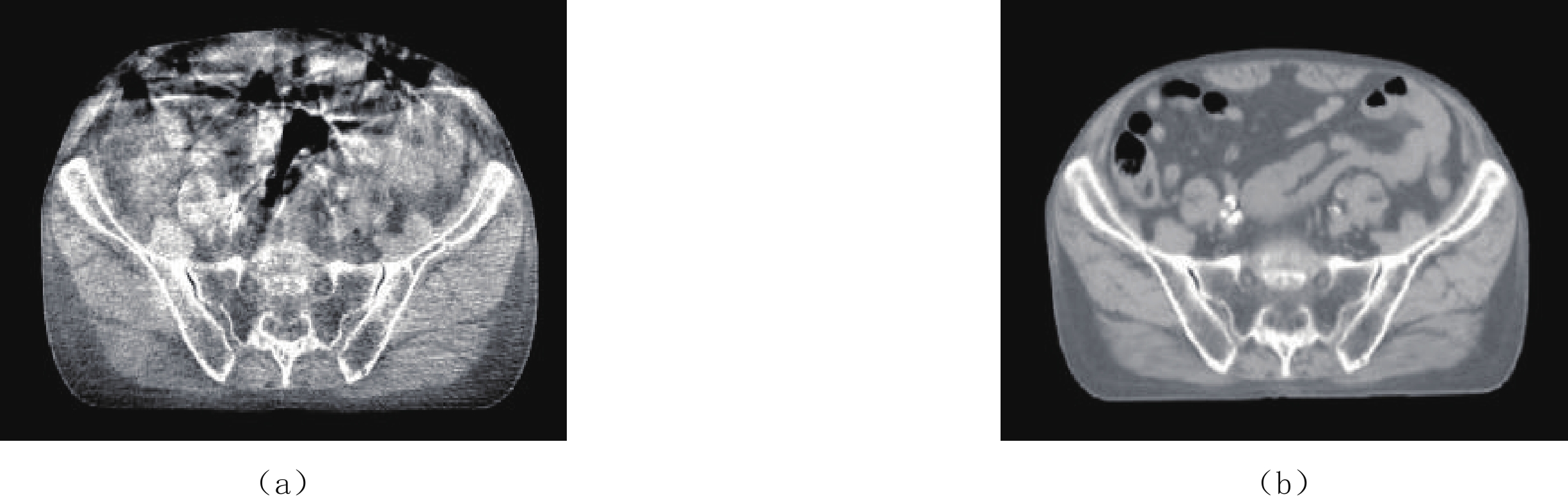
The cone-beam CT (CBCT) system based on the two-dimensional flat-panel detector technology is widely applied in patient location verification before radiotherapy. However, during the application of intraperitoneal tumor radiotherapy, severe shading and streaking artifacts caused by respiratory movement and intestinal peristalsis make it difficult to distinguish tumor areas from the CBCT images. Due to the non-rigid deformation of flexible organs such as the pancreas under the action of respiratory motion, it is hard to quantify deviation between the body surface motion monitoring results and the actual organ motion, and it is also difficult to monitor irregular motion represented by intestinal peristalsis. There is no effective solution to motion artifact correction in CBCT. Based on theory of biodynamics and common knowledge of human physiology, in this paper we propose a brand new radiotherapy image-guided cone-beam CT motion artifact correction method without motion monitoring or implantation of in-vivo markers. The proposed artifact correction strategy is designed based on the features of the artifact images and fusion of various CT image domain processing algorithms. The results suggest that the image quality of cone beam CT has been significantly improved after the application of this strategy in the clinical abdominal CBCT image processing. The average CT number error in typical soft tissue areas reduces from 90 HU to 30 HU, and the boundary of the intestinal cavity and surrounding soft tissue information are partially recovered. The proposed artifact correction strategy does not require respiratory gating or increase of projections, which can be integrated into existing workflows without marker implantation surgery. The motion-artifact-corrected CBCT images provide more accurate tumor localization information for image-guided radiotherapy of pancreatic carcinoma. The proposed method is proved practical and efficient for clinical applications
| [1] |
SIEWERDSEN J H, JAFFRAY D A. Cone-beam computed tomography with a flat-panel imager: Magnitude and effects of X-ray scatter[J]. Medical Physics, 2001, 28(2): 220−231. doi: 10.1118/1.1339879
|
| [2] |
JAFFRAY D A, SIEWERDSEN J H, WONG J W, et al. Flat-panel cone-beam computed tomography for image-guided radiation therapy[J]. International Journal of Radiation Oncology, Biology, Physics, 2002, 53(5): 1337−1349. doi: 10.1016/S0360-3016(02)02884-5
|
| [3] |
SABEL M. First results with the varian on-board imager (TM)[J]. Strahlentherapie und Onkologie, 2005: 181.
|
| [4] |
ABBAS H, CHANG B, CHEN Z J. Motion management in gastrointestinal cancers[J]. Journal of Gastrointestinal Oncology, 2014, 5(3): 223−235.
|
| [5] |
MURPHY M J, MARTIN D, WHYTE R, et al. The effectiveness of breath-holding to stabilize lung and pancreas tumors during radiosurgery[J]. International Journal of Radiation Oncology, Biology, Physics, 2002, 53(2): 475−482. doi: 10.1016/S0360-3016(01)02822-X
|
| [6] |
SONKE J J, ZIJP L, REMEIJER P, et al. Respiratory correlated cone beam CT[J]. Medical Physics, 2005, 32(4): 1176−1186. doi: 10.1118/1.1869074
|
| [7] |
LI T F, XING L, MUNRO P, et al. Four-dimensional cone-beam computed tomography using an on-board imager[J]. Medical Physics, 2006, 33(10): 3825−3833. doi: 10.1118/1.2349692
|
| [8] |
DIETRICH L, JETTER S, TUCKING T, et al. Linac-integrated 4D cone beam CT: First experimental results[J]. Physics in Medicine and Biology, 2006, 51(11): 2939−2952. doi: 10.1088/0031-9155/51/11/017
|
| [9] |
SONKE J J, ROSSI M, WOLTHAUS J, et al. Frameless stereotactic body radiotherapy for lung cancer using four-dimensional cone beam CT guidance[J]. International Journal of Radiation Oncology, Biology, Physics, 2008, 74(2): 567−574.
|
| [10] |
BISSONNETTE J P, FRANKS K N, PURDIE T G, et al. Quantifying interfraction and intrafraction tumor motion in lung stereotactic body radiotherapy using respiration-correlated cone beam computed tomography[J]. International Journal of Radiation Oncology, Biology, Physics, 2009, 75(3): 688−695. doi: 10.1016/j.ijrobp.2008.11.066
|
| [11] |
SHIEH C C, KIPRITIDIS J, O'BRIEN R T, et al. Image quality in thoracic 4D cone-beam CT: A sensitivity analysis of respiratory signal, binning method, reconstruction algorithm, and projection angular spacing[J]. Medical Physics, 2014, 41(4): 041912.
|
| [12] |
STEINER E, SHIEH C C, CAILLET V, et al. 4-Dimensional cone beam computed tomography-measured target motion underrepresents actual motion[J]. International Journal of Radiation Oncology, Biology, Physics, 2018, 102(4): 932−940. doi: 10.1016/j.ijrobp.2018.04.056
|
| [13] |
SHIMOHIGASHI Y, TOYA R, SAITO T, et al. Tumor motion changes in stereotactic body radiotherapy for liver tumors: An evaluation based on four-dimensional cone-beam computed tomography and fiducial markers[J]. Radiation Oncology, 2017, 12(1): 61.
|
| [14] |
TOYA R, SAITO T, SHIMOHIGASHI Y, et al. Four-dimensional cone-beam computed tomography-guided radiotherapy for gastric lymphoma[J]. Japanese Journal of Radiology, 2018, 36(2): 159−163. doi: 10.1007/s11604-017-0698-8
|
| [15] |
JIN P, HULSHOF M C C M, van WIERINGEN N, et al. Interfractional variability of respiration-induced esophageal tumor motion quantified using fiducial markers and four-dimensional cone-beam computed tomography[J]. Radiotherapy and Oncology, 2017, 124(1): 147−154. doi: 10.1016/j.radonc.2017.05.015
|
| [16] |
OHIRA S, ISONO M, UEDA Y, et al. Assessment with cone-beam computed tomography of intrafractional motion and interfractional position changes of resectable and borderline resectable pancreatic tumours with implanted fiducial marker[J]. British Journal of Radiology, 2017, 90(1072): 20160815.
|
| [17] |
MARCHANT T E, PRICE G J, MATUSZEWSKI B J, et al. Reduction of motion artefacts in on-board cone beam CT by warping of projection images[J]. British Journal of Radiology, 2011, 84(999): 251−264. doi: 10.1259/bjr/90983944
|
| [18] |
MARCHANT T E, SKALSKI A, MATUSZEWSKI B J. Automatic tracking of implanted fiducial markers in cone beam CT projection images[J]. Medical Physics, 2012, 39(3): 1322−1334. doi: 10.1118/1.3684959
|
| [19] |
WANG M J, SHARP G C, RIT S, et al. 2D/4D marker-free tumor tracking using 4D CBCT as the reference image[J]. Physics in Medicine and Biology, 2014, 59(9): 2219−2233. doi: 10.1088/0031-9155/59/9/2219
|
| [20] |
ZACHIU C, de SENNEVILLE B D, TIJSSEN R H N, et al. Non-rigid CT/CBCT to CBCT registration for online external beam radiotherapy guidance[J]. Physics in Medicine and Biology, 2018, 63(1): 015027.
|
| [21] |
LIU J L, ZHANG X, ZHANG X Q, et al. 5D respiratory motion model based image reconstruction algorithm for 4D cone-beam computed tomography[J]. Inverse Problem, 2015, 31(11): 115007. doi: 10.1088/0266-5611/31/11/115007
|
| [22] |
ZHANG H, MA J H, BIAN Z Y, et al. High quality 4D cone-beam CT reconstruction using motion-compensated total variation regularization[J]. Physics in Medicine and Biology, 2017, 62(8): 3313−3329. doi: 10.1088/1361-6560/aa6128
|
| [23] |
PETERLIK I, STRZELECKI A, LEHMANN M, et al. Reducing residual-motion artifacts in iterative 3D CBCT reconstruction in image-guided radiation therapy[J]. Medical Physics, 2021, 48(10): 6497−6507. doi: 10.1002/mp.15236
|
| [24] |
VOOGT J, de BOIS J, van HERK M, et al. Cone beam CT motion artifact reduction by projection image selection[C]//University of Toronto. Proceedings of the XVth International Conference on the Use of Computers in Radiation Therapy (ICCR 2007), Toronto, Canada, 2007: 13-17.
|
| [25] |
NIJKAMP J, VOOGT J, DE BOIS J, et al. Improved automatic prostate localization after cone beam CT motion artefact reduction[J]. Radiotherapy and Oncology, 2008, 88: S108−S. doi: 10.1016/j.radonc.2007.10.030
|
| [26] |
MÄKINEN Y, AZZARI L, FOI A. Collaborative filtering of correlated noise: Exact transform-domain variance for improved shrinkage and patch matching[J]. IEEE Transactions on Image Processing, 2020, 29: 8339−8354. doi: 10.1109/TIP.2020.3014721
|
| [1] | LIU Yuxin, WEI Jiaotong, ZHAO Xiaojie, CHEN Ping, PAN Jinxiao. A Correction Method for Hardening Artifacts in CT Images Based on Integral Invariance[J]. CT Theory and Applications, 2025, 34(4): 571-579. DOI: 10.15953/j.ctta.2025.064 |
| [2] | ZHANG Wenjun, HUANG Gang, DING Haining, XU Hongchun. Research Progress of Scattering Artifact Correction in Medical Cone-beam Computed Tomography Imaging Based on Deep Learning[J]. CT Theory and Applications, 2023, 32(2): 285-296. DOI: 10.15953/j.ctta.2022.131 |
| [3] | LI Zhong-chuan, MENG Fan-yong, YANG Min. The Correction of Fan-beam CT Image Artifacts Induced by Deflection of Linear-detectors[J]. CT Theory and Applications, 2014, 23(2): 237-248. |
| [4] | ZHENG Jia, ZHANG Ding-hua, HUANG Kui-dong, LI Xiang-xin. An Adaptive Enhance Method Based on Unsharp Masking for Cone Beam CT Images[J]. CT Theory and Applications, 2013, 22(2): 291-296. |
| [5] | LI Jie, YUAN Yue, ZHANG Cong-cong, YUAN Pei-xin. The Research on Match Algorithm in Tire Meridian Detection System[J]. CT Theory and Applications, 2011, 20(2): 173-181. |
| [6] | YUAN Pei-xin, ZHANG Xiao-hui. X-ray Detection of Tyre with Digital Image Processing[J]. CT Theory and Applications, 2007, 16(2): 48-51. |
| [7] | GU Jian-wei, ZHANG Li, CHEN Zhi-qiang, XING Yu-xiang, GAO He-wei. The Review of Reasons and Correction Methods for Artifacts in ICT Images[J]. CT Theory and Applications, 2005, 14(3): 24-28. |
| [8] | YUAN Pei-xin, SUN Li-na. Algorithm of Digital Image Processing for Determining an object’s True Gray Levels[J]. CT Theory and Applications, 2005, 14(2): 16-21. |
| [9] | Zu Donglin, He Qiang, Luo Qingfei, Jin Zhen, Bao Shanglian. The Correction of Motion Artifact with fMRI Using Image Registration[J]. CT Theory and Applications, 2001, 10(4): 1-3. |
| [10] | Wang Ben, Wang Ge. Image Reconstruction Algorithms for Cone-Beam CT[J]. CT Theory and Applications, 2001, 10(2): 1-8. |
Supported by: Beijing Renhe Information Technology Co. Ltd 


 DownLoad: