ISSN 1004-4140
CN 11-3017/P
| Citation: |
LU X L, LIU Y F, ZHANG Y X, et al. Advances in Measurements of Electron Density and Effective Atomic Number Using Dual-energy Computed Tomography[J]. CT Theory and Applications, 2024, 33(6): 740-746. DOI: 10.15953/j.ctta.2024.111. (in Chinese).

|
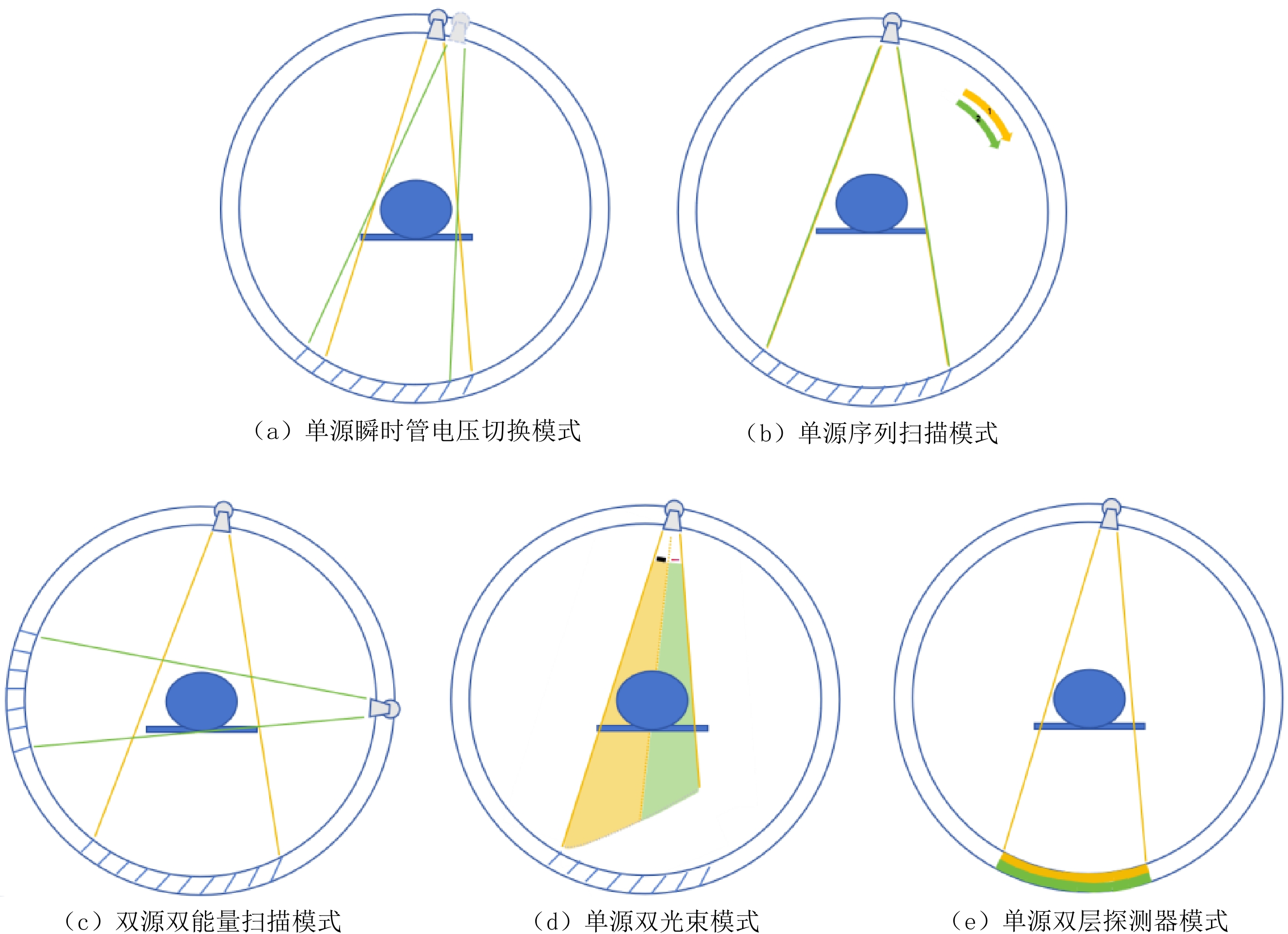
Accurate measurements of electron density and effective atomic number are essential for precise imaging diagnosis and dose estimation in the multiparametric functional analysis of dual-energy computed tomography (CT). This paper provides a review of research progress pertaining to different dual-energy imaging modalities of CT for measuring electron density and effective atomic number accurately. Its aim is to offer a reference for improving the accuracy of imaging diagnosis and treatment planning.
| [1] |
AGOSTINI A, BORGHERESI A, MARI A, et al. Dual-energy CT: Theoretical principles and clinical applications[J]. La Radiologia Medica, 2019, 124(12): 1281-1295.
|
| [2] |
PATINO M, PROCHOWSKI A, AGRAWAL M D, et al. Material separation using dual-energy CT: Current and emerging applications[J]. Radiographics, 2016, 36(4): 1087-1105.
|
| [3] |
WANG A S, HSIEH S S, PELC N J. 双能CT的基本原理、应用和未来展望[J]. CT理论与应用研究, 2012, 21(3): 367−386. (in English).
WANG A S, HSIEH S S, PELC N J. A review of dual energy CT: Principles, applications, and future outlook[J]. CT Theory and Applications, 2012, 21(3): 367−386.
|
| [4] |
OHIRA S, WASHIO H, YAGI M, et al. Estimation of electron density, effective atomic number and stopping power ratio using dual-layer computed tomography for radiotherapy treatment planning[J]. Physica Medica, 2018, 56: 34-40.
|
| [5] |
ZHAO W, SHEN S, KE T, et al. Clinical value of dual-energy CT for predicting occult metastasis in central neck lymph nodes of papillary thyroid carcinoma[J]. European Radiology, 2024, 34(1): 16-25.
|
| [6] |
ONISHI S, FUJIOKA C, KAICHI Y, et al. Utility of dual-energy CT for predicting the vascularity of meningiomas[J]. European Journal of Radiology, 2020, 123: 108790.
|
| [7] |
BHARATI A, RANI MANDAL S, GUPTA A K, et al. Non-invasive characterisation of renal stones using dual energy CT: A method to differentiate calcium stones[J]. Physica Medica, 2022, 101: 158−164.
|
| [8] |
赵聪, 刘兰平, 汤良, 等. 双肺电子密度值和放化疗模式与NSCLC放射性肺炎发生相关性[J]. 中华肿瘤防治杂志, 2017, 24(18): 1305−1309. DOI: 10.16073/j.cnki.cjcpt.2017.18.009.
ZHAO C, LIU L P, TANG L, et al. Effects of different chemotherapeutic models and double lung electron density value on the occurrence of radiation pneumonia of stage Ⅲ NSCLC[J]. Chinese Journal of Cancer Prevention and Treatment, 2017, 24(18): 1305−1309. DOI: 10.16073/j.cnki.cjcpt.2017.18.009. (in Chinese).
|
| [9] |
KONIETZKE P, STEENTOFT H H, WAGNER W L, et al. Consolidated lung on contrast-enhanced chest CT: The use of spectral-detector computed tomography parameters in differentiating atelectasis and pneumonia[J]. Heliyon, 2021, 7(5): e07066. DOI: 10.1016/j.heliyon.2021.e07066.
|
| [10] |
RUTHERFORD R A, PULLAN B R, ISHERWOOD, I. Measurement of effective atomic number and electron density using an EMI scanner[J]. Neuroradiology, 1976, 11(1): 15−21.
|
| [11] |
LI Z, RAVISHANKAR S, LONG Y, et al. Learned mixed material models for efficient clustering based dual-energy CT image decomposition[C]//2018 IEEE Global Conference on Signal and Information Processing (GlobalSIP). IEEE, 2018: 529-533.
|
| [12] |
郭俏, 姚旭峰. 双能CT图像域基材料分解算法的研究进展[J]. CT理论与应用研究, 2023, 32(1): 139−146. DOI: 10.15953/j.ctta.2021.067.
GUO Q, YAO X F. Progress of material decomposition algorithms in dual-energy CT imaging[J]. CT Theory and Applications, 2023, 32(1): 139−146. DOI: 10.15953/j.ctta.2021.067. (in Chinese).
|
| [13] |
BROOKS R A, Di CHIRO G. Beam hardening in X-ray reconstructive tomography[J]. Physics in Medicine & Biology, 1976, 21(3): 390−398.
|
| [14] |
SCHAEFFER C J, LEON S M, OLGUIN C A, et al. Accuracy and reproducibility of effective atomic number and electron density measurements from sequential dual energy CT[J]. Medical Physics, 2021, 48(7): 3525−3539.
|
| [15] |
ALMEDIDA I P, SCHYNS L E, ÖLLERS M C, et al. Dual-energy CT quantitative imaging: A comparison study between twin-beam and dual-source CT scanners[J]. Medical Physics, 2017, 44(1): 171-179.
|
| [16] |
HUA C H, SHAPIRA N, MERCHANT T E, et al. Accuracy of electron density, effective atomic number, and iodine concentration determination with a dual-layer dual-energy computed tomography system[J]. Medical Physics, 2018, 45(6): 2486−2497.
|
| [17] |
LANDRY G, GAUDREAULT M, Van ELMPT W, et al. Improved dose calculation accuracy for low energy brachytherapy by optimizing dual energy CT imaging protocols for noise reduction using sinogram affirmed iterative reconstruction[J]. Zeitschrift für Medizinische Physik, 2016, 26(1): 75-87.
|
| [18] |
RGEL A, BIER G, HENNERSDORF F, et al. Image quality of CT angiography of supra-aortic arteries: Comparison between advanced modelled iterative reconstruction (ADMIRE), sinogram affirmed iterative reconstruction (SAFIRE) and filtered back projection (FBP) in one patients' group[J]. Clinical Neuroradiology, 2020, (1). DOI: 10.1007/s00062-018-0740-y.
|
| [19] |
徐同江, 王旭, 李艳, 等. IMR、iDose4和FBP三种重建算法在隆鼻整形术CT扫描的应用研究[J]. CT理论与应用研究, 2022, 31(3): 357−364. DOI: 10.15953/j.ctta.2021.072.
XU T J, WANG X, LI Y, et al. Application of IMR, iDose4 and FBP reconstruction algorithms in CT scanning of rhinoplasty[J]. CT Theory and Applications, 2022, 31(3): 357−364. DOI: 10.15953/j.ctta.2021.072. (in Chinese).
|
| [20] |
DE M P, ORIGGI D. New adaptive statistical iterative reconstruction ASiR-V: Assessment of noise performance in comparison to ASiR[J]. Journal of Applied Clinical Medical Physics, 2018, 19(2): 275−286. DOI: 10.1002/acm2.12253.
|
| [21] |
田璐, 黄红云, 范杰. 双能CT螺距和迭代重建权重对单能衰减和有效原子序数的影响: 体模研究[J]. 中国医学影像技术, 2023, 39(1): 99−103. DOI: 10.13929/j.issn.1003-3289.2023.01.023.
TIAN L, HUANG H Y, FAN J. Impact of pitch and iterative reconstruction weight on monochromatic attenuation and effective atomic number of dual-energy CT: A phantom study[J]. Chinese Journal of Medical Imaging Technology, 2023, 39(1): 99−103. DOI: 10.13929/j.issn.1003-3289.2023.01.023. (in Chinese).
|
| [22] |
中华医学会放射学分会, 中国医师协会放射医师分会, 安徽省影像临床医学研究中心. 能量CT临床应用中国专家共识[J]. 中华放射学杂志, 2022, 56(5): 476−487.
|
| [23] |
ALKADHI H, EULER A, MAINTZ D, et al. Spectral imaging: Dual-energy, multi-energy and photon-counting CT[M]. Cham: Springer International Publishing, 2022.
|
| [24] |
GOODSITT M M, CHRISTODOULOU E G, LARSON S C. Accuracies of the synthesized monochromatic CT numbers and effective atomic numbers obtained with a rapid kVp switching dual energy CT scanner[J]. Medical Physics, 2011, 38(4): 2222−2232. (in Chinese).
|
| [25] |
KAWAHARA D, OZAWA S, YOKOMACHI K, et al. Synthesized effective atomic numbers for commercially available dual-energy CT[J]. Reports of Practical Oncology & Radiotherapy, 2020, 25(4): 692−697.
|
| [26] |
OGATA T, UEGUCHI T, YAGI M, et al. Feasibility and accuracy of relative electron density determined by virtual monochromatic CT value subtraction at two different energies using the gemstone spectral imaging[J]. Radiation Oncology, 2013, 8: 83.
|
| [27] |
TATSUGAMI F, HIGAKI T, KIGUCHI M, et al. Measurement of electron density and effective atomic number by dual-energy scan using a 320-detector computed tomography scanner with raw data-based analysis: A phantom study[J]. Journal of Computer Assisted Tomography, 2014, 38(6): 824−827.
|
| [28] |
KAWAHARA D, OZAWA S, YOKOMACHI K, et al. Accuracy of the raw-data-based effective atomic numbers and monochromatic CT numbers for contrast medium with a dual-energy CT technique[J]. The British Journal of Radiology, 2018, 91(1082): 20170524.
|
| [29] |
KAWAHARA D, OZAWA S, TANAKA S, et al. Automatic contrast medium extraction system using electron density data with dual-energy CT[J]. The British Journal of Radiology, 2018, 91(1090): 20180396.
|
| [30] |
MCCOLLOUGH C H, LENG S, YU L, et al. Dual- and multi-energy CT: Principles, technical approaches, and clinical applications[J]. Radiology, 2015, 276(3): 637−653.
|
| [31] |
王鹏朝, 单春辉, 郭方凯, 等. 不同双能量组合对Rho/Z检测准确性的模体研究[J]. 中国医疗设备, 2021. DOI: 10.3969/j.issn.1674-1633.2021.09.016.
WANG P C, SHAN C H, GUO F K, et al. Phantom study on the accuracy of Rho/Z detection with different dual energy combinations[J]. China Medical Devices, 2021. DOI:10.3969/j.issn.1674-1633.2021.09.016(in Chinese).
|
| [32] |
LANDRY G, RENIERS B, GRANTON P V, et al. Extracting atomic numbers and electron densities from a dual source dual energy CT scanner: Experiments and a simulation model[J]. Radiotherapy and Oncology, 2011, 100(3): 375−379.
|
| [33] |
LANDRY G, DORRINGER F, SI-MOHAMED S, et al. Technical note: Relative proton stopping power estimation from virtual monoenergetic images reconstructed from dual-layer computed tomography[J]. Medical Physics, 2019, 46(4): 1821−1828.
|
| [34] |
YANG M, WOHLFAHRT P, SHEN C, et al. Dual- and multi-energy CT for particle stopping-power estimation: Current state, challenges and potential[J]. Physics in Medicine & Biology, 2023, 68(4). DOI: 10.1088/1361-6560/acabfa.
|
| 1. |
刘雨欣,陈凡秀,孙洁,王远,王潇,于洋,顾焱吉. 管电压管电流和开机时间对CT图像质量的影响. 实验力学. 2024(01): 17-26 .

| |
| 2. |
朱丽娟,马瑞,沈云,汪芳,杨彦兵,曹永佩,杨利莉,吴小红. 探讨不同管电压下DLIR重建算法对冠状动脉CTA图像质量的影响. 宁夏医学杂志. 2023(09): 787-791+865 .
| |
| 3. |
王振宇,黄蕾. 分析CT图像的质量控制与设备维护保养. 中国设备工程. 2023(18): 85-87 .
|
Supported by: Beijing Renhe Information Technology Co. Ltd 


 DownLoad: